Analyze how experimental results and claims are presented, scoped, and limited in a paper.Know what the science actually supports before you trust the answer.
Press Enter ↵ to analyze
Explore by Goal
"Science is organized knowledge. Wisdom is organized life."
- Immanuel Kant
Quick Explanation
Copied
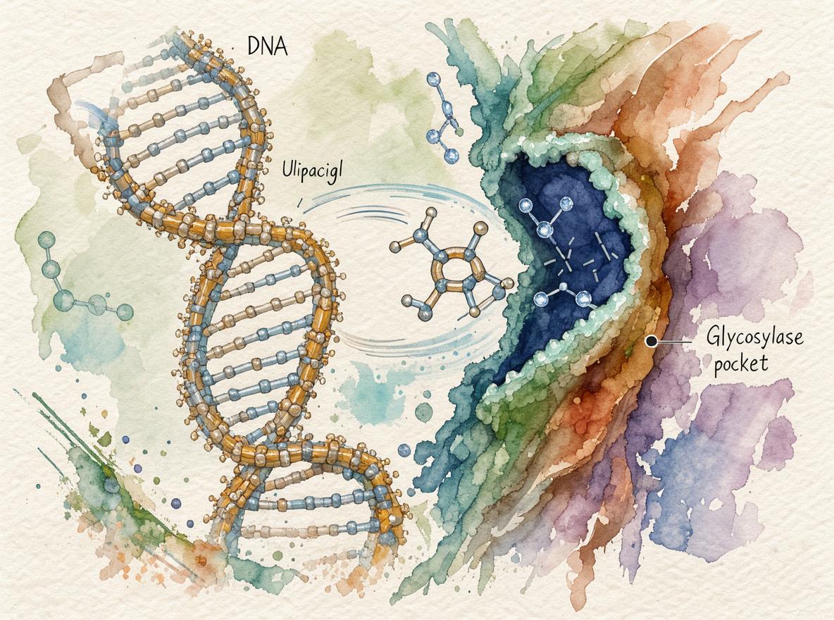
Parikh, Putnam & Tainer (2000) integrate crystal structures of human, HSV-1, and E. coli UDG into a coherent 'pinch–push–pull' mechanism: minor-groove backbone compression detects uracil, loop-mediated flipping drives catalysis via a dissociative SN1-like reaction, and product AP-sites are handed off to APE1 .
Long Explanation
The review's central achievement is converting ~20 PDB structures (1AKZ, 1SSP, 1EMH, 1UGH, etc., 1.40–3.20 Å) into a functional mechanism: (1) processive minor-groove scanning with ~4 Å phosphate compression and ~45° DNA bending, (2) leucine-intercalation-driven uracil flipping, (3) dissociative SN1-like glycosylic bond cleavage coupling anomeric and σ–π stereoelectronic effects, and (4) AP-site retention with hand-off to APE1 . Binding kinetics (KD 48 nM; kass 7.9×10⁶ M⁻¹s⁻¹) from the companion EMBO study anchor the two-step scanning/flipping model .
Interpretation vs. observation. The pinch–push–pull mechanism is an author inference from static structures plus fluorescence data; the authors themselves flag two open questions—whether UDG flips every nucleotide during scanning, and the quantitative contribution of the dissociative pathway to bond cleavage . Later work partially validated the mechanism: MD simulations show mechanical deformation aids uracil recognition in dsDNA , but also show that within nucleosomes only a fraction of uracils are accessible —a blindspot the review does not address, since chromatin impairs glycosylase access as shown earlier . All evidence is in vitro/crystallographic; in vivo hand-off dynamics remain inferred, and generalization to other glycosylases is argued, not demonstrated.
Feedback:
Updated: November 01, 2026
BGPT Paper Review
Study Novelty
80%
For 2000, the synthesis of pinch–push–pull scanning, coupled stereoelectronic catalysis, and APE1 hand-off was a major conceptual advance; the ideas built on the authors' own 1998 structural work, so novelty is high but not wholly new.
Scientific Quality
90%
Mechanistic claims are tightly tied to high-resolution structures (1.4–3.2 Å) and kinetics; limitations: static snapshots, in vitro scope, and author-acknowledged untested scanning assumptions.
Study Generality
90%
Explicitly frames UDG principles (minor-groove scanning, base flipping, AP-site hand-off) as general to DNA glycosylases, supported by later UNG2/SMUG1 and archaeal PCNA studies.
Study Usefulness
90%
Framework guided two decades of glycosylase research, inhibitor design (e.g., HSV-1 OctAU), and chromatin-context follow-ups; core teaching reference for BER initiation.
Study Reproducibility
80%
All 21 PDB entries are tabulated with resolutions and references, enabling direct re-analysis; some kinetic and figure details rely on companion papers.
Explanatory Depth
90%
Mechanistic depth is exceptional—from atomic stereoelectronic coupling (anomeric + σ–π effects) to cellular pathway coordination—though the scanning step remains semi-quantitative.
Retrieving and aligning the 21 listed UDG/Ugi PDB structures to compare active-site geometry and interface residues across human, HSV, and E. coli enzymes.
Get emailed when your analysis is done!
We'll email you the results when your analysis is finished.
Hypothesis Graveyard
Serendipitous capture of spontaneously flipped-out bases: rejected because backbone compression precedes flipping per fluorescence two-step kinetics and Ugi mimicry of pre-flipped DNA.

 Explore by Goal
 Quick Explanation
 Long Explanation
 Top Data Sources
 Ask a Follow-Up
 Key Insight
 Keep Exploring
 Analysis
 Hypothesis Graveyard
 Potential Experiments
 Discussion
