Check each claim against its experiments, reported results, scope, and stated limitations.Know what the science actually supports before you trust the answer.
Press Enter ↵ to start review
Explore by Goal
"The important thing is not to stop questioning. Curiosity has its own reason for existing."
- Albert Einstein
Quick Explanation
Copied
CD34-derived human Langerhans cells (LCs) fail to phosphorylate ERK after TLR2/3/7/8 stimulation, secrete no IL-12p70, and drive Th2-biased T-cell responses, whereas monocyte-derived DCs (moDCs) phosphorylate ERK, secrete IL-12p70, and skew toward Th1 — showing subset-specific TLR signalling is largely post-transcriptional and ERK-independent
Long Explanation
Core findings
Duraisingham et al. compared in vitro CD34+-derived LCs (35–40% langerin+CD1a+HLA-DR+) with moDCs across TLR agonists, signalling inhibitors, phospho-ERK flow cytometry, T-cell co-cultures, and an 84-gene TLR PCR array . The central, surprising observation: LCs — the most efficient Th2 inducers — show no ERK phosphorylation within 60 min of TLR2, 3 or 7/8 stimulation, contradicting the ERK→Th2 model established in TLR2-stimulated moDCs.
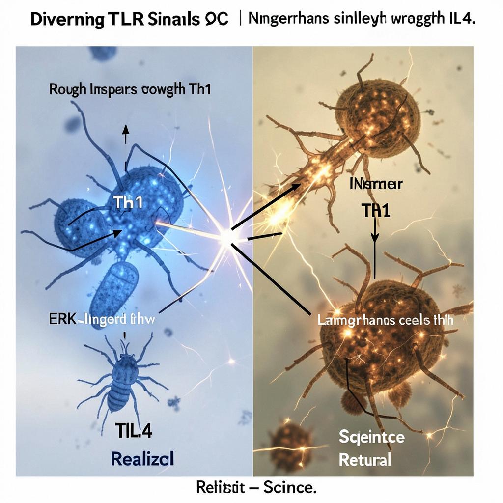
Schematic of the paper's key qualitative result (Figure 3). PMA did phosphorylate ERK in LCs, ruling out detection failure.
Supporting evidence
Cytokines: LCs secreted IL-12/23p40 only after TLR7/8 and never IL-12p70; moDCs produced IL-12p70 after TLR3/4/7/8
T-cell skewing: even unstimulated LCs induced IL-4+ T cells (amplified by TLR2-LPS); moDCs drove IFN-γ+ Th1 responses regardless of agonist
Inhibitors: p38 inhibition suppressed moDC maturation (CD83/HLA-DR) but only LC CD83; PI3K inhibition reduced moDC survival but increased LC CD83 — divergent pathway usage
Transcriptomics: of 84 genes, major differences lay in TLRs themselves (LC: TLR1/7/10 high; moDC: TLR3/8 high) and cytokines (LC: IL-1/TNF high; moDC: IL-10 high), plus LCs expressing 6-fold more MD-1 and 3-fold more RP105 — negative TLR4 regulators explaining their LPS unresponsiveness
Reported fold-differences (approximate, from Figures 5 and Table S1; blue = higher in LCs, orange = higher in moDCs).
Critical appraisal
Strengths: clean paired design (six independent experiments for FACS/ELISA; five for p-ERK), a PMA positive control proving LC ERK competence, two-fold inhibitor dose testing, and gDNA contamination checks on the PCR array .
Blind spots and limits: (1) LCs were only 35–40% langerin+, and T-cell data came from magnetically purified CD1a+ cells from just three co-culture experiments — small n with donor-pooling that can mask inter-individual variation. (2) Pharmacological inhibitors (SB203580, SP600125, FR180204, LY294002, SN50) have known off-targets; no siRNA/CRISPR confirmation was attempted. (3) The ERK null result rests on a 60-min window — later kinetics or phospho-proteomics were not explored. (4) In vitro CD34-derived LCs may not equal bona fide epidermal LCs, and skin-isolated LCs reportedly lack TLR4 mRNA entirely . (5) The mechanistic claim of "constitutive, ERK-independent Th2 induction" is an inference — the actual IL-4-driving signal in LCs remains unidentified. No in vivo validation was attempted.
What would falsify the conclusion: p-ERK induction in LCs beyond 60 min, intact ERK-dependent Th2 skewing in primary skin LCs, or genetically (not pharmacologically) confirmed p38/PI3K dependency of LC maturation.
Context: this was published in 2010, when the Langerhans-cell-paradigm debate (LCs as tolerogenic vs immunogenic) was active; the Th2-bias finding aligns with Klechevsky's earlier subset-specialization work. Modern single-cell RNA-seq and phospho-proteomics could resolve whether the post-transcriptional hypothesis holds.
Feedback:
Updated: October 02, 2026
BGPT Paper Review
Study Novelty
90%
First demonstration that human LCs do not phosphorylate ERK upon TLR stimulation, contradicting the ERK→Th2 dogma from moDC studies; subset-specific TLR signaling in human LCs was largely unexplored.
Scientific Quality
90%
Well-controlled experiments (PMA control, 6 replicates, inhibitor dose-testing), rigorous stats, no conflicts, MRC funding; but small n for T-cell co-cultures, reliance on pharmacological inhibitors, and in vitro LC model limit certainty.
Study Generality
60%
Findings are specific to two human in vitro DC subsets and a limited agonist set, but the principle that same-TLR stimulation produces subset-specific signaling has broad immunological relevance.
Study Usefulness
80%
Directly informs skin immunology, vaccine/adjuvant design for skin-targeted delivery, and explains Th2 skewing at mucosal/barrier sites; molecular pathway divergence matters for DC-based immunotherapy.
Study Reproducibility
70%
Detailed methods, defined agonist/inhibitor concentrations, and six independent experiments; but no data repositories, pooled PCR-array samples, and modest sample sizes hamper exact replication.
Explanatory Depth
80%
Integrates phenotype, cytokine, T-cell skewing, signaling, and transcriptomics into a coherent subset-difference model; falls short of identifying the actual ERK-independent Th2 mechanism.
SIMULATED model of ERK phosphorylation kinetics in moDCs (induced) vs LCs (flat), based on the paper's reported timepoints and qualitative finding that LCs show no p-ERK within 60 min. Assumes baseline = 1, moDC peak ~3-fold at 60 min, SE = 20% of signal. NOT observed data.
Generated scientific data; not direct experimental measurements.
Get emailed when your analysis is done!
We'll email you the results when your analysis is finished.
Hypothesis Graveyard
ERK phosphorylation is required for Th2 induction in all human DC subsets — falsified by CL097 (ERK+ but Th1) and LC TLR2-LPS (Th2 without ERK) in the same paper.
TLR stimulation produces identical signaling regardless of DC subset — falsified by divergent p38/JNK/PI3K inhibitor sensitivity and p-ERK outcomes between LCs and moDCs.

 Explore by Goal
 Quick Explanation
 Long Explanation
 Top Data Sources
 Ask a Follow-Up
 Key Insight
 Keep Exploring
 DataGen
 Hypothesis Graveyard
 Discussion
