Full-text extraction of experimental results, figures, and citations for fast, transparent reviews.
Press Enter ↵ to solve
Fuel Your Discoveries
"The day science begins to study non-physical phenomena, it will make more progress in one decade than in all the previous centuries of its existence."
- Nikola Tesla
Quick Answer
Copied
Sox17 acts as an arterial-identity gatekeeper
In this 2013 Nature Communications study, endothelial loss of Sox17 causes failure to acquire/maintain arterial programs (arteries gain venous markers), disrupts vascular remodeling, and yields embryonic lethality (embryo) or retinal hypersprouting with arteriovenous malformations (postnatal retina). Mechanistically, the paper supports a Wnt/β-catenin → Sox17 → Notch regulatory linkage via direct promoter binding to Notch4 and Dll4 (strong evidence within their experimental system).
Primary paper:
Long Answer
Paper Review (visual-first): Sox17 is indispensable for acquisition and maintenance of arterial identity
What it claims (core result): endothelial Sox17 is arterial-restricted and required for arterial identity acquisition and maintenance; Sox17 loss drives arterial→venous marker switching, abnormal sprouting, and (depending on timing/model) embryonic lethality or retinal arteriovenous malformations. The paper’s mechanistic claim is that Sox17 functions upstream of Notch and is regulated by canonical Wnt/β-catenin signaling, forming a Wnt→Sox17→Notch axis.
Evidence type mix (from this paper): in vivo conditional genetics + retinal whole-mount phenotyping + qRT-PCR marker profiling + pharmacology (DAPT) + genetic Notch perturbations + venous-to-arterial transcriptional conversion by Sox17 overexpression + ChIP promoter binding evidence + lineage/marker co-localization.
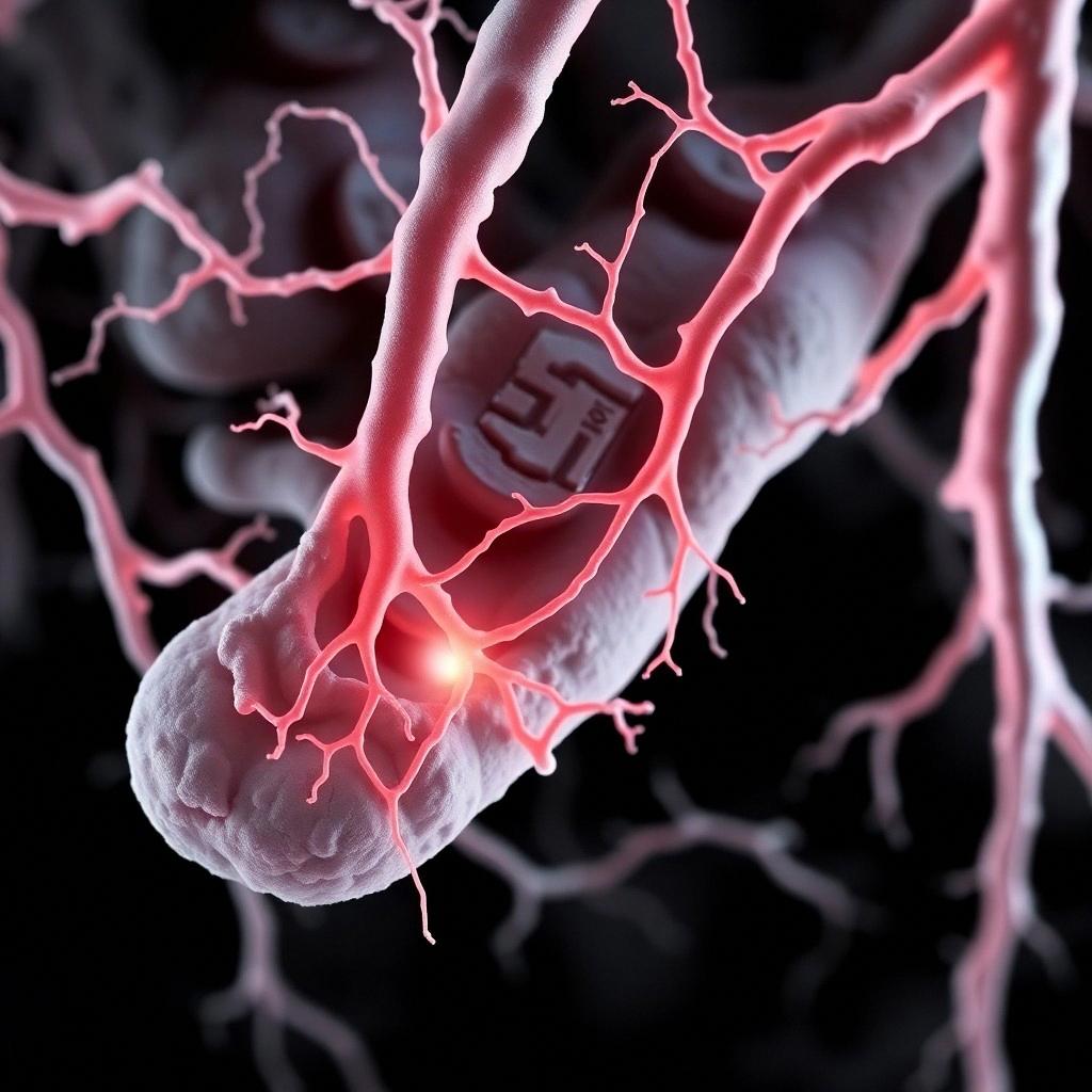
1) Key visualization: Sox17 arterial restriction and magnitude
The paper quantifies representative nuclear fluorescence intensities in P5 retina: ~200 ± 30 a.u. in arterial endothelium vs ~50 ± 10 a.u. in veins/capillaries (n≈100 nuclei/group; exact details stated in the text).
The paper proposes: canonical Wnt/β-catenin upregulates Sox17, and Sox17 then induces Notch pathway activity, promoting arterial differentiation; Sox17 inhibition/ablation prevents Notch activation and arterial identity acquisition.
3) Phenotype map: what changes when Sox17 is endothelial-deleted?
Embryo (Tie2-Cre; Sox17 ECKO): endothelial deletion causes in utero lethality (100%) between E10.5–E12.5, with major vascular remodeling defects and failure of large artery development; arterial/venous differentiation is disrupted.
Postnatal retina (Cdh5(PAC)-CreER; tamoxifen P1): Sox17 ablation leads to hypersprouting (including multidirectional sprouting), reduced vascular progression, dynamic tip-cell filopodia behavior consistent with impaired inhibitory Notch influences, reduced arterial differentiation markers, increased venous markers, and large arteriovenous malformations.
4) Causality logic test: is Sox17 upstream or downstream of Notch?
The paper tests Notch→Sox17 and Sox17→Notch directionality in vivo:
Notch inhibition (DAPT) does not change Sox17 expression, while it downregulates Notch target genes (Hes/Hey1/Hey2).
Notch activation (Notch IC gain-of-function in endothelium) does not significantly change Sox17 expression.
Sox17 gain-of-function in venous endothelial cells increases NICD nuclear translocation and upregulates Notch-pathway genes (including Hey1, Dll4, Dll1, Notch4) and arterial markers; and Sox17 promoter binding to Notch4 and Dll4 is supported by ChIP-qPCR in their cellular contexts.
5) Skeptical critique: what could weaken the interpretation?
(A) Genetic background / redundancy confound: The authors discuss discrepancy with a prior global Sox17 deletion study, attributing it to genetic background and possible functional redundancy among SoxF family members.
(B) Off-target or pathway-crosstalk for pharmacology: DAPT inhibits Notch signaling but can have off-target effects or influence other pathways indirectly. The paper uses multiple Notch perturbations (pharmacologic and genetic) to mitigate this, but residual uncertainty remains because Notch pathway is pleiotropic and retinal developmental programs are interconnected.
(C) Binding vs functional necessity: ChIP demonstrates Sox17 binding to Notch4/Dll4 promoter regions, but the paper (as provided here) does not show promoter/enhancer mutagenesis that abolishes Sox17 binding while preserving overall protein levels. Thus, binding is consistent with causality but not fully definitive about necessity of those exact sites.
(D) Species and context generalization: The strongest mechanistic evidence is in mouse endothelial contexts and cultured venous endothelial manipulations. Translating the exact Wnt→Sox17→Notch wiring to human arterial biology requires caution; multiple later studies on Sox17 in vascular disease suggest context-dependent roles (e.g., aneurysm under hypertension).
(E) Publication/interpretation bias check via corroboration: Independent reports can help triangulate. For example, Notch-related work has been connected to Sox17’s angiogenic regulation in other contexts, supporting some directionality/functional involvement.
6) What would most change my confidence (disproof points)
If Sox17 were dispensable for arterial marker acquisition/maintenance under improved genetic background controls (or if strong phenotypes were attributable to off-target effects of the Tie2-Cre / Cdh5-CreER drivers), the “indispensable” framing would weaken.
If Wnt/β-catenin manipulations did not reliably shift Sox17 expression in endothelial contexts, or if Sox17 could be removed without affecting Notch target genes and arterial differentiation (i.e., Sox17 is correlated but not causal), then the regulatory axis would be incorrect.
If enhancer/promoter disruption of the reported Sox17-binding sites within Notch4/Dll4 left overall Sox17 function intact and failed to recapitulate loss-of-function phenotypes, it would indicate those bindings are not sufficient/necessary. (This is an uncertainty because such site-specific mutagenesis isn’t shown in the provided excerpt.)
Author-review deep dives (jump links)
Click to open BGPT author reviews for the paper’s authors.
Need deeper analysis (optional agent)?
Run an AI Scientist agent to extract additional quantitative details from the full-text and cross-check pathway logic with other Sox17 vascular papers.
Feedback:
Updated: March 24, 2026
BGPT Paper Review
Study Novelty
90%
The work introduces Sox17 as an arterial-identity transcription factor with direct upstream control over Notch pathway components and positions it mechanistically in a Wnt→Sox17→Notch axis for arterial/venous specification, supported by endothelial-specific loss/gain-of-function and ChIP evidence in mouse vascular contexts.
Scientific Quality
80%
Strong causal triangulation in vivo (endothelial-specific conditional KO at multiple developmental stages) plus mechanistic directionality tests (Notch perturbations vs Sox17 perturbations), marker switching, and ChIP-binding evidence. Main weaknesses are that promoter/enhancer necessity for the specific Sox17 binding sites is not fully established in the excerpt, and the ChIP-binding evidence is not the same as genetic disruption of the sites; also, Cre-driver specificity and pharmacologic off-targets remain general concerns.
Study Generality
70%
Arterial/venous identity is broadly conserved across vertebrate vasculatures, but the strongest evidence is mouse endothelial development/retina; the exact Wnt→Sox17→Notch wiring may vary by tissue and species context. The paper itself flags disease relevance as needing clarification and notes context differences (e.g., tumor vasculature, strain modifiers).
Study Usefulness
90%
Mechanistic framework and experimentally tractable Sox17–Notch–Wnt integration provides a roadmap for future studies on arterial identity maintenance and its dysregulation in vascular malformations and related disease models.
Study Reproducibility
80%
Methods include defined mouse strains, Cre lines, tamoxifen timing, DAPT administration, and experimental assays (qRT-PCR, ChIP-qPCR, whole-mount immunostaining), with quantification described and statistical testing reported. Remaining reproducibility uncertainty: the excerpt does not list raw data accessions or complete n for every panel.
Explanatory Depth
90%
Beyond correlation, it attempts mechanistic directionality (Notch→Sox17 vs Sox17→Notch), shows transcriptional conversion-like effects of Sox17 in venous endothelial cells, and provides direct binding evidence to Notch4/Dll4 promoter regions; together these support a deeper causal network explanation.
Extract Sox17-related marker genes and Notch/Wnt pathway targets from the paper text, then generate a small gene-network table mapping each perturbation (Sox17 KO/overexpression, Notch inhibition) to direction of change.
Get emailed when your analysis is done!
We'll email you the results when your analysis is finished.
Hypothesis Graveyard
“Sox17 phenotypes are primarily a generic developmental delay/artifact from Cre toxicity.” The paper reports consistent arterial-to-venous marker switching and Notch axis changes tied to Sox17 perturbations across embryonic vs postnatal retina contexts, which argues against a purely nonspecific delay explanation.
“Notch is downstream of Sox17 only indirectly via VEGF.” The paper reports that VEGF stimulation/upregulation did not significantly change Sox17 expression and that Sox17 upregulation does not follow VEGF activation in their endothelial systems, arguing against a simple VEGF→Sox17 link as the primary driver.