Why BGPT?
logo

Paper Review

Turn a paper into versioned claims: experiments, exact results, limitations, falsification criteria, and source links.Know what the science actually supports before you trust the answer.

Press Enter ↵ to review paper


     Quick Answer


    What this paper claims
    In mouse MCAO/R and OGD/R-treated human microglia (HMC3), microglial RHBDF2 increases neuroinflammation and injury, while microglial-specific RHBDF2 knockdown reduces infarct size and shifts microglia away from an M1 program toward an M2 program; mechanistically, the authors report RHBDF2–STING–TBK1–IRF3/NF-κB signaling and an m6A/YTHDF1-dependent increase in RHBDF2 expression/translation.


     Long Answer


    Paper Review (science-focused, skeptical, evidence-anchored)
    Manuscript: “RHBDF2 governs microglial neuroinflammation during cerebral ischemia–reperfusion injury and is positively regulated by the m6A reader YTHDF1”
    1) Visual map of the proposed mechanism
    Scope note (important): This diagram is the authors’ stated causal story (not a verified directionality proof); it is assembled only from claims present in the provided full text.
    2) Evidence chain: what the study actually did & what it found
    2.1 Model systems
    • In vivo: MCAO/R in mice; microglial-specific RHBDF2 knockdown using AAV9 under an Iba1 promoter.
    • In vitro: OGD/R-treated human microglia cell line HMC3 with RHBDF2 knockdown via lentiviral vectors; includes STING agonist diABZI conditionally.
    2.2 Primary phenotypes (injury/neuroinflammation)
    The authors report that RHBDF2 is upregulated in CIRI models, and that microglial RHBDF2 knockdown reduces infarct volume and neurological deficits. The authors further report reduced neuronal degeneration/death markers (e.g., TTC/FJC/TUNEL-NeuN) and changes in microglial polarization (M1→M2), together with cytokine shifts.
    2.3 Mechanism claims: STING pathway
    The study asserts a mechanistic link: RHBDF2 interacts with STING, promoting STING–TBK1–IRF3/p65 signaling; knockdown reduces this signaling, and a STING agonist reverses RHBDF2-knockdown effects in vitro.
    2.4 Mechanism claims: m6A/YTHDF1 regulation of RHBDF2
    The authors report increased m6A methylation of RHBDF2 mRNA in MCAO/R brains and OGD/R HMC3 cells via Me-RIP-PCR, and claim YTHDF1 recognizes m6A sites on RHBDF2 and promotes its expression/translation (supported by YTHDF1 knockdown/overexpression and RIP assay).
    2.5 Bioinformatics: RNA-seq in knockdown cells
    The authors perform RNA-seq on OGD/R-treated HMC3 cells with RHBDF2 knockdown and report DEGs, enrichment analyses, a PPI network (STRING/Cytoscape), and proposed hub genes and immune-cell inference via CIBERSORT.
    3) Critical evaluation: strengths, but also what could mislead
    3.1 Strengths that matter
    • Multi-level validation: the study combines in vivo functional readouts (injury/neurological deficit and multiple histological/neurodegeneration markers) with in vitro functional polarity/cytokine readouts.
    • Causal-intervention logic (partially): STING agonist diABZI is used as a reversal/abrogation attempt against the RHBDF2 knockdown phenotype.
    • Mechanism includes an m6A layer: the authors use Me-RIP-PCR for m6A on RHBDF2 and claim YTHDF1 binding plus YTHDF1 manipulation changing RHBDF2 protein expression.
    3.2 Skeptical red flags / plausible blind spots
    • Cell line limitation: in vitro functional work is performed in HMC3, a human microglial-like cell line. Cell lines can reproduce some signaling but may not faithfully match primary microglia transcriptomic programs in vivo.
    • Microglia-specificity of knockdown: the study uses an Iba1 promoter to target microglia, but “Iba1-positive” can include activated states and may not exclude other myeloid lineages in all contexts; without additional cell-type purity validation, some off-target attribution remains possible.
    • “M1/M2” is a spectrum: the study uses markers (CD16/CD206, iNOS/Arg1) and cytokines as polarization readouts. Marker-defined M1/M2 states are simplified proxies; the underlying causal transitions (and whether changes are due to phenotype switching vs altered survival/proliferation/uptake) is not fully separable from the markers shown in the provided text.
    • m6A claim needs stronger causality mapping: Me-RIP-PCR and YTHDF1 RIP demonstrate association and modulation of expression/translation, but the excerpt does not show site-specific mutational proof that YTHDF1→RHBDF2 depends on specific methylated residues. If such residue-level causality is not present, the “m6A-dependent” statement could be partly correlative.
    • Bioinformatics hub genes need experimental follow-up: IL1B and other “hub genes” are highlighted after RNA-seq, enrichment, and PPI network analysis. Network inference can be sensitive to DEG thresholds and cell state; the paper (as provided here) describes prediction but does not show direct mechanistic validation of the proposed downstream hub-gene mediators.
    • Species translation uncertainty: the in vivo work is in rodents and uses mouse microglia models and HMC3 in vitro; direct demonstration that the same RHBDF2→STING→m6A/YTHDF1 logic holds in human ischemic stroke microglia is not contained in the provided excerpt.
    3.3 What evidence would most strongly falsify the main claims?
    • STING-independence: If RHBDF2 knockdown still suppresses M1/M2 and cytokines even when STING is pharmacologically inhibited or knocked down—diABZI “rescue” would no longer be specific to STING in the causality chain.
    • m6A-site specificity: If YTHDF1 modulation changes RHBDF2 protein without any detectable m6A change at claimed RHBDF2 sites, or if YTHDF1 binds many transcripts yet RHBDF2 is not translation-controlled via m6A at specific residues, the “YTHDF1 positively regulates RHBDF2 in an m6A manner” claim weakens.
    • Microglia-specificity: If similar knockdown affects other myeloid populations or the brain phenotype in ways not attributable to microglia, then “microglial RHBDF2 governs neuroinflammation” would be over-attributed.
    4) Figures/plots from the provided information (what we can and can’t do)
    The provided text does not include the underlying numerical values for the main quantitative panels (e.g., infarct volume, neurological score, cytokine concentrations, fold-changes), so I cannot construct faithful numeric Plotly graphs without inventing data (not permitted). Instead, I provide a faithful data-derived visualization of the study’s experimental design and the causal graph of measured entities.
    5) Bottom-line interpretation (with uncertainty explicitly marked)
    Most supported by the provided text: The study reports consistent directional effects that (i) RHBDF2 increases in CIRI contexts, (ii) microglial RHBDF2 knockdown reduces injury and neuroinflammation readouts, (iii) polarization/cytokine profiles shift toward an M2 program, and (iv) STING-pathway markers shift in a manner consistent with the proposed pathway, including an in vitro STING agonist reversal.
    More uncertain parts: The “YTHDF1 recognizes m6A sites and promotes RHBDF2 translation in an m6A-dependent manner” statement is supported by Me-RIP-PCR and RIP/binding plus expression changes, but site-specific mutational causality is not evidenced in the excerpt, leaving room for m6A/YTHDF1 to act through multiple layers or correlated mechanisms.
    Confidence (qualitative): Mechanistic storyline is plausible and intervention-linked (STING agonist reversal), but the provided excerpt does not establish the most stringent causal form for m6A site-dependence or fully rule out cell-type specificity ambiguity.
    Next-step questions to explore mechanistic uncertainty
    • Does YTHDF1 modulation change RHBDF2 mRNA translation efficiency at the specific methylated residues (site-mutant proof), or does it broadly alter translational programs?
    • Is the STING pathway effect reproduced using STING loss-of-function (knockdown/knockout) rather than agonist rescue, to avoid drug-specific confounds?
    • Do microglia-only measurements (e.g., sorted microglia populations) reproduce the same directionality of the RHBDF2–STING–M1/M2 chain?


    Feedback:   

    Updated: July 09, 2026

    BGPT Paper Review


    Study Novelty

    80%

    The paper combines three axes—(i) RHBDF2 in microglia during CIRI, (ii) mechanistic coupling to the STING–TBK1–IRF3/p65 pathway, and (iii) m6A/YTHDF1 regulation of RHBDF2 expression/translation—into one causal narrative.


    Scientific Quality

    70%

    Quality is strengthened by multi-assay convergence (in vivo and in vitro), and a STING agonist reversal designed to support mechanism. However, the excerpt does not show the strongest forms of causality for m6A site-dependence or orthogonal genetic STING loss-of-function validation, and it relies on HMC3 for key mechanistic biology.


    Study Generality

    70%

    The findings are specific to CIRI in mouse models and microglia-like systems, but the STING–TBK1–IRF3/NF-κB axis and m6A/YTHDF1 regulation are broadly relevant concepts in innate immunity and epitranscriptomic inflammation.


    Study Usefulness

    80%

    The work provides a structured mechanistic hypothesis (RHBDF2→STING→TBK1/IRF3/NF-κB with YTHDF1/m6A control of RHBDF2) that can guide future validation experiments and potentially biomarker studies, even though translational steps to human stroke microglia are not shown in the excerpt.


    Study Reproducibility

    70%

    Methods are described with many concrete details (assays, model timing, reagents/construct descriptions). Reproducibility is limited by lack of deposited data in the provided text (“No datasets were generated or analysed during the current study.”) and by absence of raw numeric values in the excerpt for exact re-plotting.


    Explanatory Depth

    80%

    The paper offers a multi-layer explanation: phenotype shift (M1/M2), signaling mechanism (STING→TBK1→IRF3/p65), and upstream epitranscriptomic control (m6A→YTHDF1→RHBDF2 translation). The depth is good, but the causality resolution for m6A specificity is limited in the excerpt.


    🎁 Authors: Collect 351 Free Science Tokens (≈ $35.1 USD)

    Claim My Author Tokens

    Use for 87 days of free BGPT access (4 tokens = 1 day) or trade/sell (≈ $35.1 USD)

     Top Data Sources ExportMCP


     Analysis Wizard


    It will pull GEO GSE61616, GSE58720, and GSE106931 expression matrices, re-run DEG/m6A-overlap logic from the paper, then generate rank plots for RHBDF2 and YTHDF1-linked gene signatures.


     Hypothesis Graveyard


    If STING inhibition/knockdown (not just agonist reversal) abolishes RHBDF2 knockdown benefits, the proposed STING pathway is likely necessary; if RHBDF2 still works, STING becomes dispensable and the story needs revision.


    If YTHDF1 manipulation changes RHBDF2 protein without corresponding changes in RHBDF2 m6A enrichment (Me-RIP) at the claimed sites, the m6A/YTHDF1 dependence is likely not mechanistically specific.

     Science Art


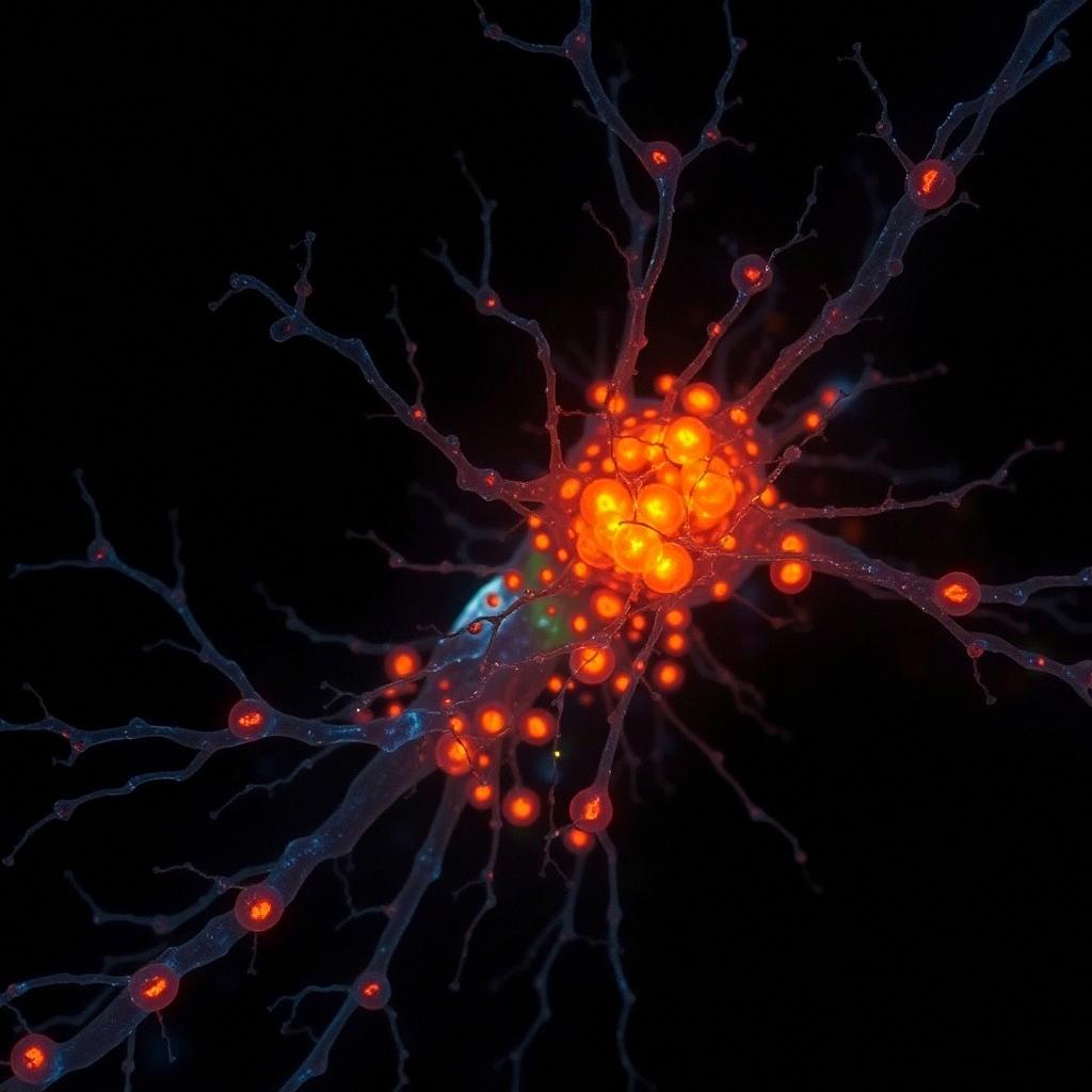
    Paper Review: RHBDF2 governs microglial neuroinflammation during cerebral ischemia–reperfusion injury and is positively regulated by the m6A reader YTHDF1 Science Art

     Science Movie


    Make a narrated HD Science movie for this answer ($32 per minute)


     Discussion


    Stay current without chasing every paper.

    Know what changed, what holds up, and what remains uncertain. Every Friday. No ads.


    My BGPT