Ask questions across claims linked to experiments, exact results, limitations, and sources.Know what falsifiable science actually supports before you trust the answer.
Press Enter ↵ to search evidence
Explore by Goal
"Biology is a science of three dimensions. The first is the study of each species across all levels of biological organization, molecule to cell to organism to population to ecosystem. The second dimension is the diversity of all species in the biosphere. The third dimension is the history of each species in turn, comprising both its genetic evolution and the environmental change that drove the evolution."
- E. O. Wilson
Quick Answer
Copied
This preprint overturns the prevailing model that LEDGF's H3K36me3-binding PWWP domain determines HIV-1 integration site selection, showing instead that MLL1 tethers LEDGF to TSSs via the IBD to recruit RNA Pol II, while integration occurs across gene bodies through a proposed IBD handoff during elongation. The core evidence is strong genome-wide ChIP-seq showing 96% of LEDGF peaks lost upon MLL1 knockdown and no change in gene-level integration upon SETD2/H3K36me3 loss. However, the proposed competition mechanism remains inferential and is demonstrated only in HEK293T cells.
Long Answer
Core Evidence: MLL1, Not H3K36me3, Positions LEDGF at Promoters
Using CRISPR-engineered biallelic 3XFLAG tagging of endogenous LEDGF in HEK293T cells, the authors obtained high-specificity ChIP-seq maps revealing that LEDGF is concentrated at transcription start sites (TSSs) — 95% of LEDGF-3XFLAG peaks overlapped H3K4me3 and 54% overlapped RNA Pol II (MACS2, FDR<0.05). Critically, 96% of LEDGF-3XFLAG peaks were significantly reduced upon shRNA knockdown of MLL1 (KMT2A), while deleting the IBD domain reduced 81% of LEDGF peaks. Conversely, 69% of MLL1 peaks overlapping LEDGF were reduced in LEDGF knockout cells, indicating reciprocal stabilization . These data are consistent with prior structural work showing the LEDGF IBD binds an MLL1 intrinsically disordered motif in a menin-dependent complex .
H3K36me3 Is Dispensible for Gene-Level Integration Specificity
The most provocative negative result: SETD2 knockout (eliminating all H3K36me3) did not significantly alter integration frequencies per gene, recurrent integration gene (RIG) overlap coefficient was unchanged (no significant change), and SPIN-state preferences were unaltered. Only a modest upstream shift in the metagene integration profile was observed, consistent with H3K36me3 contributing to distribution within gene bodies but not to gene selection . This contradicts prior biochemical and structural evidence that the PWWP domain specifically recognizes H3K36me3-nucleosomes . The dPWWP construct retained strong TSS enrichment, while MLL1 knockdown did not recapitulate LEDGF knockout integration phenotypes (RIG overlap coefficient 0.95 vs. control), suggesting residual LEDGF at TSSs in MLL1-depleted cells remains sufficient for integration positioning.
The Dual-Role Model: Strengths and Critical Gaps
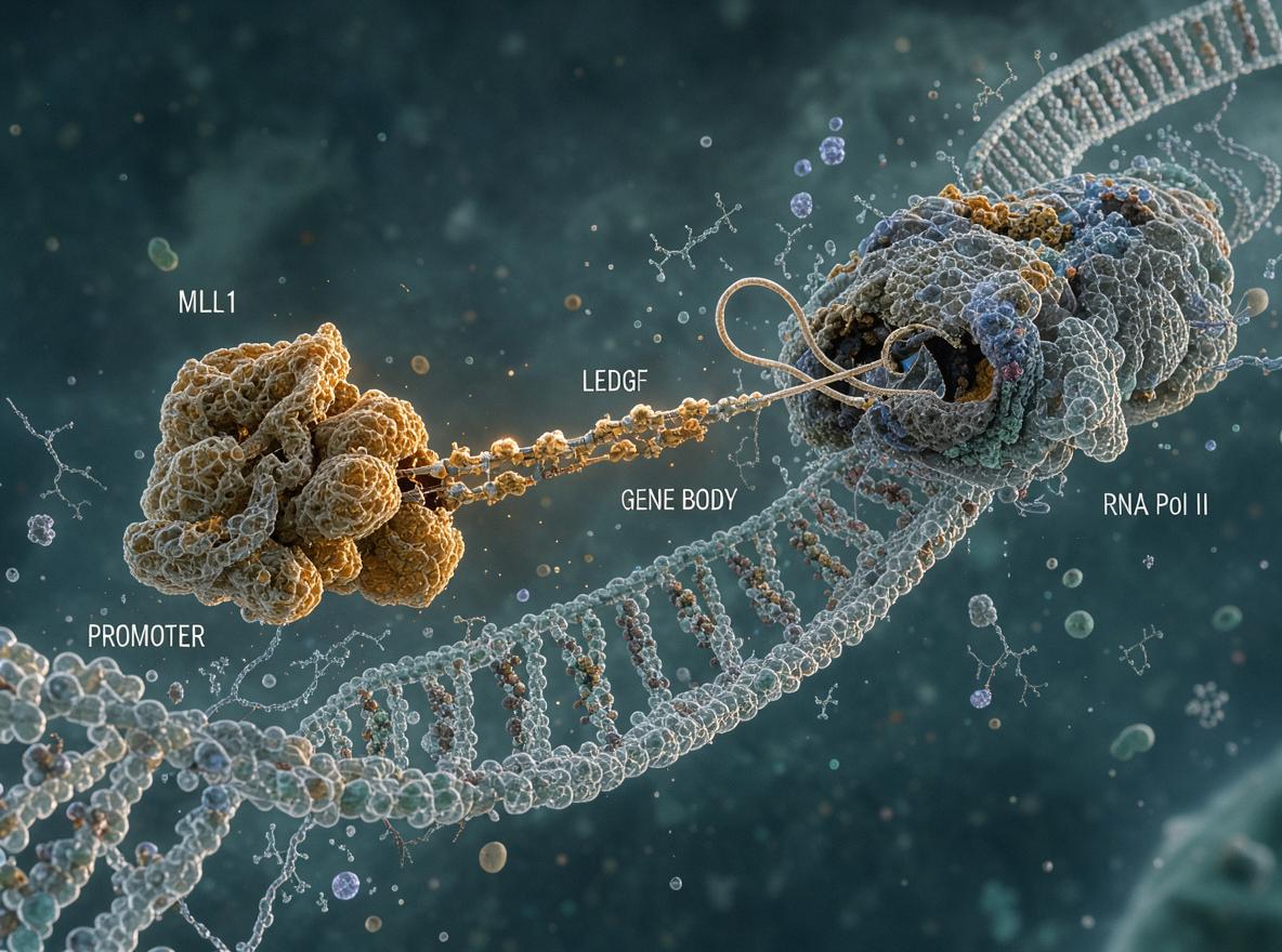
The proposed model is elegant: at TSSs, MLL1 occupies the IBD, blocking IN access; during elongation, LEDGF transfers to elongation factors (IWS1, SPT6, TFIIS), freeing the IBD for IN binding and enabling integration across transcribed sequences . Evidence for this includes: (1) HOX genes show strong LEDGF/MLL1 co-enrichment across gene bodies but low integration; (2) IBD-deleted LEDGF loses TSS binding; (3) MLL1 KD does not alter integration patterns. However, no direct biochemical competition assay in cells demonstrates that MLL1 outcompetes IN for the IBD at TSSs. Prior structural work showed HIV-1 IN can outcompete MLL from the IBD in vitro (apparent IC₅₀ 39.7 nM) , which seems to contradict the authors' in-cell inference unless chromatin context, stoichiometry, or co-factor recruitment reverses this affinity relationship.
Additional context from related work: INLAIs targeting the LEDGF-binding pocket on IN confirm that disrupting this interface is antivirally relevant , and R-loop formation has been independently proposed as an additional integration determinant .
Limitations and Blind Spots
The study is confined to HEK293T cells — an embryonic kidney-derived line not representative of primary CD4+ T cells, the physiologically relevant HIV-1 target. ChIP-seq of endogenous 3XFLAG-LEDGF required ectopic rescue constructs expressed from Sleeping Beauty vectors (with 260,991 peaks called, suggesting substantial noise: only 1.7% overlapped endogenous LEDGF peaks). MLL1 was knocked down by shRNA (not genetically deleted), leaving open residual-protein explanations. The MLL1-blocking-IN mechanism remains untested in cells. Cross-linking-based ChIP may artifactually trap transient interactions, potentially explaining the TSS-enrichment pattern that differs from native-chromatin methods reporting more even LEDGF distribution .
Author Reviews:
Feedback:
Updated: November 19, 2026
BGPT Paper Review
Study Novelty
70%
Challenges the dominant H3K36me3-centric model of LEDGF chromatin targeting with genome-wide ChIP-seq evidence and proposes a novel MLL1-dependent promoter-tethering mechanism. The dual-role model is conceptually new but builds on established MLL1-IBD structural knowledge and prior elongation-factor interactions.
Scientific Quality
70%
Well-controlled with multiple genetic perturbations (LEDGF KO, dPWWP, IBD deletion, MLL1 KD, SETD2 KO) and orthogonal readouts. Major weaknesses: single cell line (HEK293T), ectopic expression with high noise (1.7% peak overlap), shRNA rather than genetic MLL1 deletion, no direct biochemical competition assay in cells to support the central MLL1-blocks-IN inference, and cross-linking artifacts acknowledged but not controlled for. Not yet peer-reviewed.
Study Generality
60%
Findings are demonstrated only in HEK293T cells. Whether LEDGF TSS enrichment and MLL1-dependence hold in primary CD4+ T cells, macrophages, or other physiologically relevant HIV-1 target cells remains unknown. The MLL1-leukemia connection broadens relevance but the core HIV-1 integration mechanism may be cell-type-specific.
Study Usefulness
80%
Directly informs HIV-1 integration site selection biology, identifies MLL1 as a gatekeeper preventing integration at TSSs, provides mechanistic context for MLL1-fusion leukemia dependence on LEDGF, and suggests new therapeutic angles targeting the MLL1-IBD interface. The H3K36me3-negative result redirects the field away from a widely held but apparently incorrect assumption.
Study Reproducibility
70%
Detailed methods with specific reagents, software versions, and parameters are provided. CRISPR-engineered cell lines are well-characterized. However, single-lab, single-cell-type data without independent replication; the 3XFLAG-LEDGF ectopic rescue produced 260,991 peaks (high noise), raising questions about signal-to-noise in that dataset.
We'll email you the results when your analysis is finished.
Hypothesis Graveyard
H3K36me3-PWWP interaction as the primary determinant of HIV-1 integration gene specificity: falsified by SETD2 KO data showing no change in per-gene integration frequencies, RIG overlap, or SPIN-state preferences. H3K36me3 contributes only to intra-gene distribution, not gene selection.
LEDGF-dependent integration being driven by direct PWWP-H3K36me3 recognition of target chromatin: contradicted by the finding that dPWWP LEDGF retains strong TSS enrichment and that integration profiles across gene bodies are largely unchanged in SETD2 KO cells.

 Explore by Goal
 Quick Answer
 Long Answer
 Top Data Sources
 Ask a Follow-Up
 Key Insight
 Keep Exploring
 Hypothesis Graveyard
 Potential Experiments
 Discussion
