Check each claim against its linked experiments, results, and falsification criteria.Know what the science actually supports before you trust the answer.
Press Enter ↵ to check claims
Explore by Goal
"The most exciting phrase to hear in science, the one that heralds new discoveries, is not 'Eureka!' but 'That's funny...'"
- Isaac Asimov
Quick Answer
Copied
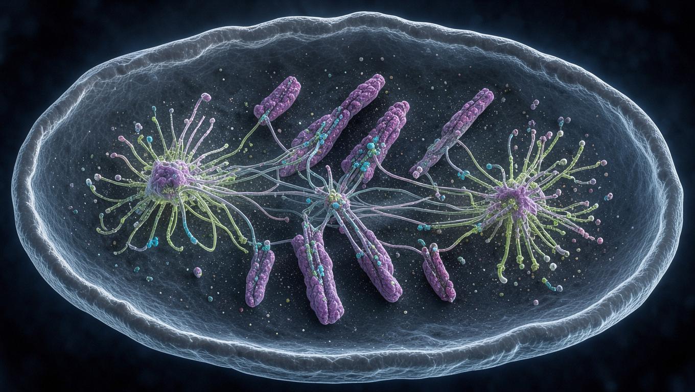
Dai et al. (2009) show that haspin RNAi in U2OS/HeLa cells causes ectopic γ-tubulin foci and spindle disarray, and live-cell imaging plus rescue experiments (topoisomerase II inhibition, monastrol) argue these defects are indirect consequences of lost chromosome cohesion rather than direct centrosomal failure .
Long Answer
Core Evidence: Cohesion Loss, Not Centrosome Failure
Haspin siRNA reduced H3T3ph in 76% of mitotic U2OS cells with extensive chromosome misalignment, and 67% of mitotic cells contained ≥3 γ-tubulin foci versus only 8% of controls (92% of controls had exactly two foci) . Several design choices strengthen the causal interpretation: (1) extra foci appeared in mitosis, not interphase/prophase, with no centrosome overamplification and weaker γ-tubulin intensity (~⅓ of true centrosomes); (2) live imaging showed foci emerged only after chromosomes lost metaphase-like alignment; (3) Scc1 and Sgo1 depletion phenocopied the effect, with >4 foci rising from 6% (control) to 44% (haspin), 41% (Scc1) and 28% (Sgo1) .
Two perturbation experiments are the strongest causal evidence: ICRF-193 topoisomerase II inhibition (preserving chromatid association via catenation) reduced ≥3 foci in haspin-depleted but not control cells, and kinesin-5 inhibition with monastrol both prevented formation and reversed pre-existing ectopic foci — live imaging showed foci fusing back onto dominant poles within 40 minutes, with 66% of haspin-depleted cells regaining multi-foci within 20 minutes of washout . Timing data support a sequential model: congression took 19±3 min (control), 28±12 min (haspin group I) and 46±23 min (haspin group II); group II cells then stalled at a metaphase-like stage for 1–4 hours before foci appeared .
Model and Critique
The authors argue cohesion loss disrupts integration of chromosome- and centrosome-initiated microtubule nucleation pathways, with kinesin-5 driving chromatin-nucleated microtubules into dynamic ectopic poles . Limitations: two cancer-derived cell lines only; siRNA incomplete-depletion artifacts are acknowledged but not quantified; ICRF-193 rescue is partial and drug-toxicity caveats exist; direct centrosomal haspin functions are not excluded (EGFP-haspin localizes to poles); no biochemical mechanism links cohesion loss to PCM redistribution. The mechanistic framing is an interpretation, not a directly demonstrated pathway. Later work places haspin under TOP2A-SUMOylation control at centromeres , consistent with haspin's cohesion-linked, chromosome-centered role rather than a centrosomal one. What would change the conclusion: a cohesion-independent haspin mutant rescuing poles, or centrosome defects preceding misalignment in clonal, fully-depleted cells.
Feedback:
Updated: October 31, 2026
BGPT Paper Review
Study Novelty
70%
Reinterpretation of a long-standing assumption (cohesin proteins acting at centrosomes) by showing indirect effects; careful epistasis-style rescue logic was comparatively novel for 2009 but built on prior Scc1/Sgo1 spindle-defect reports.
Scientific Quality
80%
Multiple orthogonal approaches (fixed imaging, live imaging, drug rescue, phenocopy across three cohesion factors, regrowth assays) with statistics; weaknesses include reliance on two cancer cell lines, partial rescue, unquantified knockdown depth, and no direct test of residual haspin at poles.
Study Generality
70%
Conclusions generalize beyond haspin to any cohesion-compromising manipulation (Scc1, Sgo1 phenocopies), but are cell-line specific and mitosis-focused.
Study Usefulness
70%
Useful caution for the field when interpreting centrosome defects after cohesion perturbations, and mechanistic groundwork relevant to chromosomal instability in cancers and haspin inhibitor design.
Study Reproducibility
70%
Detailed siRNA sequences, antibody dilutions, imaging parameters and cell lines are reported; raw data and n per condition partially reported (e.g., 23–116 cells in Fig. 5), but no deposited datasets.
Explanatory Depth
70%
Strong correlation-plus-rescue logic and a coherent model, but the molecular mechanism linking cohesion loss to γ-tubulin redistribution remains inferred rather than demonstrated.
We'll email you the results when your analysis is finished.
Hypothesis Graveyard
Direct centrosomal haspin function causes spindle defects: rejected (provisionally) because foci appear only after cohesion loss, are microtubule- and kinesin-5-dependent, and phenocopy Scc1/Sgo1 depletion; a local role is not fully excluded.
Centrosome overamplification causes multipolarity: ruled out by unchanged interphase/prophase foci counts, normal centriole numbers, and low γ-tubulin intensity of ectopic foci.

 Explore by Goal
 Quick Answer
 Long Answer
 Top Data Sources
 Ask a Follow-Up
 Key Insight
 Keep Exploring
 Hypothesis Graveyard
 Potential Experiments
 Discussion
