Analyze how experimental results and claims are presented, scoped, and limited in a paper.Know what the science actually supports before you trust the answer.
Press Enter ↵ to analyze
Explore by Goal
"The science of today is the technology of tomorrow."
- Edward Teller
Quick Explanation
Copied
Harris et al. (PNAS 2012) built a substrate-free tensile testing device for suspended MDCK-II epithelial monolayers, reporting tissue stiffness of 20 ± 2 kPa, >69% strain at failure, and ~1.7 μN intercellular separation forces ninefold above doublet measurements, implicating actin, myosin contractility, cadherin adhesion, and keratin filaments in monolayer mechanics. The work is a rigorous, well-controlled methods innovation whose main limits are a single cell line, ~10 μm assumed thickness in stress normalization, and short observation timescales.
Long Explanation
A first direct measurement of suspended epithelial mechanics
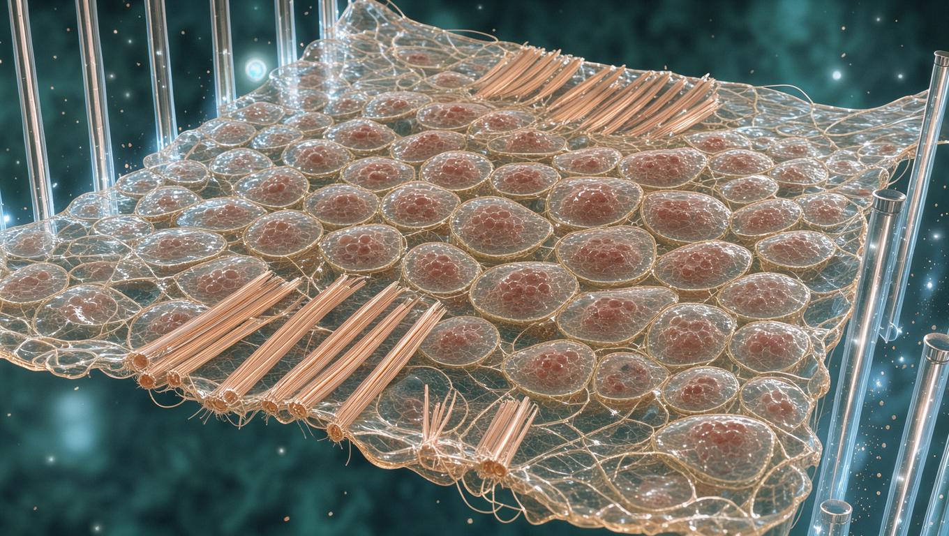
The paper's central contribution is a novel two-rod tensile testing device: monolayers are cultured on a sacrificial collagen scaffold between NiTi wires, released by collagenase digestion, and stretched while force is read from cantilever deflection calibrated against PDMS standards . This design satisfies four stated requirements — load borne only by the monolayer, minimal sample manipulation, live imaging during stretch, and quantitative force readout — a genuinely clever solution to the confounding that plagues substrate-attached measurements.
Quantitative findings, with uncertainty
Tissue stiffness was E = 20 ± 2 kPa (N = 12 controls), roughly 100-fold above transverse AFM measurements of constituent cells; monolayers tolerated 69 ± 14% strain before failing, mostly by rod delamination. Nick experiments forced intercellular rupture, yielding adhesion energy Γ ≈ 0.07 N/m and a per-cell separation force of ~1.7 μN versus ~200 nN for doublets contacted only ~30 min — evidence that junctions mature toward far stronger coupling in confluent tissue . Perturbations cleanly parsed contributors: latrunculin B halved stiffness (10 ± 6 kPa), Rho-kinase inhibition reduced it 36% (13 ± 6 kPa), and EDTA cadherin disruption collapsed it to 0.8 ± 0.4 kPa (N = 8–12 per group) .
Mechanism: shape change, not intercalation
Texture correlation showed tissue strain (εxx = 50 ± 6%) matched cellular strain measured from segmented E-cadherin-GFP images, with conserved cell volume and transverse thinning — no intercalation on ~5 min timescales. Myosin regulatory light chain relocalized to junctions upon substrate removal, and keratin filaments transitioned from wavy to taut under high strain, supporting a nonlinear load-bearing role consistent with prior keratin network work . High-stress creep showed power-law behavior (β = 0.15 ± 0.03, below single-cell values of ~0.3–0.5) with a stress threshold separating solid-like from fluid-like response.
Assumptions for the simulated curve: linear region slope fixed at 20 kPa, toe extends to ~25% strain, plateau near failure, per reported qualitative features; values are illustrative, not measured.
Critical assessment and blind spots
Strengths include device calibration against independent PDMS measurements, live/dead and polarity controls confirming monolayer health for >3 h post-digestion, and strain-rate insensitivity confirming quasi-static loading. Key limitations: stress normalization assumes uniform ~10 μm monolayer thickness; failure is dominated by cell–rod adhesion, so doublet-force estimates require a fracture-mechanics model with assumed cell diameter (a ≈ 10 μm); only MDCK-II is tested, so generality across epithelia is asserted, not shown; sample sizes are modest (N = 8–12); and the keratin mechanistic claim remains correlative — the authors themselves note it "will necessitate further study." The later literature the paper seeded — collective-migration and fluidization modeling and mechanochemical homeostasis work on the same MDCK system — confirms the paper's lasting influence. What would change the conclusions: intercalation-dominated extension at longer timescales, or keratin-disruption experiments showing no effect on high-strain stiffness.
Feedback:
Updated: November 01, 2026
BGPT Paper Review
Study Novelty
90%
First direct tensile measurement of substrate-free mature monolayers at tissue, cellular, and subcellular scales; the device and the 9-fold junction maturation force result were unprecedented.
Scientific Quality
90%
Calibrated force measurement, multiple internal controls (PDMS validation, viability/polarity checks, strain-rate tests), and clear statistics. Main gaps: single cell line, assumed uniform 10 μm thickness for stress, correlative-only keratin claims, modest N for perturbations.
Study Generality
80%
Claims apply broadly to any strongly junction-coupled epithelium, but only MDCK-II was tested; developmental extrapolations remain inferential.
Study Usefulness
90%
Provides directly usable device designs, calibration protocols, and parameter values (E, Γ, viscosity) that later modeling and tissue-engineering work adopted.
Study Reproducibility
80%
Detailed SI fabrication, calibration, and image-analysis methods support replication, though custom hardware and MATLAB code are not publicly deposited.
Explanatory Depth
90%
Multiscale linkage from molecular perturbations to tissue-level stiffness with quantitative fracture-mechanics framework and explicit falsification criteria.
Compiling and visualizing reported MDCK monolayer stiffness, failure strain, and perturbation datasets to support quantitative comparison and modeling of epithelial tissue mechanics.
Get emailed when your analysis is done!
We'll email you the results when your analysis is finished.
Hypothesis Graveyard
Monolayer mechanics are simply single-cell mechanics summed in parallel — falsified by the 100-fold stiffness mismatch between tissue and transversally probed cells.
Tissue extension requires cell intercalation as in morphogenesis — falsified by matching tissue and cellular strain tensors over 5-min timescales.

 Explore by Goal
 Quick Explanation
 Long Explanation
 Top Data Sources
 Ask a Follow-Up
 Key Insight
 Keep Exploring
 Analysis
 Hypothesis Graveyard
 Potential Experiments
 Discussion
