Ask questions across claims linked to experiments, exact results, limitations, and sources.Know what the science actually supports before you trust the answer.
Press Enter ↵ to search evidence
Explore by Goal
"Science is the great antidote to the poison of enthusiasm and superstition."
- Adam Smith
Long Answer
Copied
Nanobanana prompt
Create a publication-quality graphical abstract for an ACS Nano submission in a strict horizontal 9:4 aspect ratio, corresponding to 9 cm × 4 cm. Use a clean white background with a very subtle blue hexagonal or molecular motif. Render a scientifically precise, elegant 3D illustration with crisp vector-like outlines, restrained depth of field, and a coordinated palette of deep navy blue, cyan, orange, green, and red. Use only short, readable, sans-serif labels; avoid decorative text, unsupported quantitative claims, and visual clutter.
Left: engineered siRNA polyplex. Show a large, clearly magnified cross-section of a spherical polyplex. Depict a compact positively charged polymeric core containing multiple folded siRNA duplexes, with a distinct fluorescent tracking dye shown as small luminous green molecules. Illustrate a hydrated PEG corona as flexible blue chains extending from the surface and a small number of orange cyclic-peptide targeting ligands. Label the components exactly: “siRNA cargo,” “fluorescent tracer,” “PEG corona,” and “targeting ligand.” The particle architecture should be visually consistent with a nucleic-acid nanocarrier, while avoiding invented chemical structures or claims about a specific polymer composition. Fluorescent labeling should be shown as a tracking method, not as therapeutic activity. Evidence that intracellular siRNA release from endosomes is important for knockdown is reported in HeLa-cell experiments, but the precise polyplex architecture requested here is not established by the supplied studies .
Center: circulation and tumor access. Show several smaller copies of the same nanoplex moving through a stylized blood vessel containing red blood cells and a thin endothelial lining. Use a rightward directional arrow toward a tumor region with irregular, leaky microvessels. Illustrate limited extravasation through endothelial gaps and local particle accumulation, but label this region conservatively as “Systemic delivery and tumor accumulation.” Do not present the enhanced permeability and retention effect as universal or guaranteed; do not draw uniform accumulation throughout the tumor. The supplied evidence includes nanoparticle uptake and delivery studies, but does not directly demonstrate systemic tumor accumulation, EPR-mediated transport, or this exact siRNA formulation in vivo. Keep those elements as a conceptual pathway rather than a reported result .
Right: cancer-cell mechanism. Enlarge one cancer cell and show: receptor engagement at the plasma membrane, receptor-mediated endocytosis, endosomal maturation, endosomal escape, cytoplasmic siRNA release, RISC-mediated target-mRNA cleavage or destabilization, reduced target-protein output, and an apoptosis icon showing membrane blebbing and fragmented nuclear DNA. Label the sequence with concise captions: “Receptor binding,” “Endocytosis,” “Endosomal escape,” “Cytoplasmic siRNA release,” “Gene silencing,” and “Apoptotic response.” Use “EGFR” only if EGFR targeting and expression are experimentally established for the specific study; otherwise label the receptor “target receptor.” Use “gene silencing” only as the intended mechanistic endpoint unless knockdown data are available. The supplied siRNA study supports the importance of cytosolic release for knockdown, whereas the supplied targeted-cisplatin study supports receptor-mediated uptake only for a different HER2-targeted carrier and payload .
Composition and layout. Arrange the narrative strictly from left to right: particle design → bloodstream transport → tumor access → cancer-cell internalization → intracellular release → gene silencing → apoptosis. Use one dominant continuous arrow with smaller curved arrows only where necessary. Keep all labels inside the 9:4 canvas, with generous margins and no overlapping text. Use consistent particle size within each scene, but allow the left particle and right cell to be enlarged for explanatory clarity. Use blue arrows for transport, orange arrows for targeting, green signals for fluorescence, and red signals for apoptosis. Do not include numerical efficacy values, claims of clinical translation, or a legend unless supplied experimental data justify them. Export as a high-resolution, lossless image suitable for scientific layout, with clean edges and legible text at final 9 cm × 4 cm size.
Scientific accuracy safeguards: distinguish the intended mechanism from demonstrated evidence; do not imply that EPR is reliable in every tumor; do not imply that fluorescence proves intact siRNA release; do not infer apoptosis from gene-silencing imagery without apoptosis measurements; do not combine results from unrelated dextran, magnetic, cisplatin, insulin, and mitochondrial-delivery systems into one validated therapeutic platform. The supplied studies demonstrate nanoparticle uptake, controlled delivery, receptor-targeting precedents, and siRNA endosomal-release principles, but they do not collectively validate this exact multifunctional tumor polyplex .
We'll email you the results when your analysis is finished.
Hypothesis Graveyard
A generic EPR icon should not be treated as evidence that this nanoparticle will accumulate effectively in every tumor, because the supplied records do not provide tumor biodistribution or comparative EPR measurements.
Fluorescent intracellular signal should not be interpreted as proof of functional siRNA release or gene silencing; fluorescence can remain associated with intact particles or endosomal compartments.

 Explore by Goal
 Long Answer
 Top Data Sources
 Ask a Follow-Up
 Key Insight
 Keep Exploring
 Hypothesis Graveyard
 Potential Experiments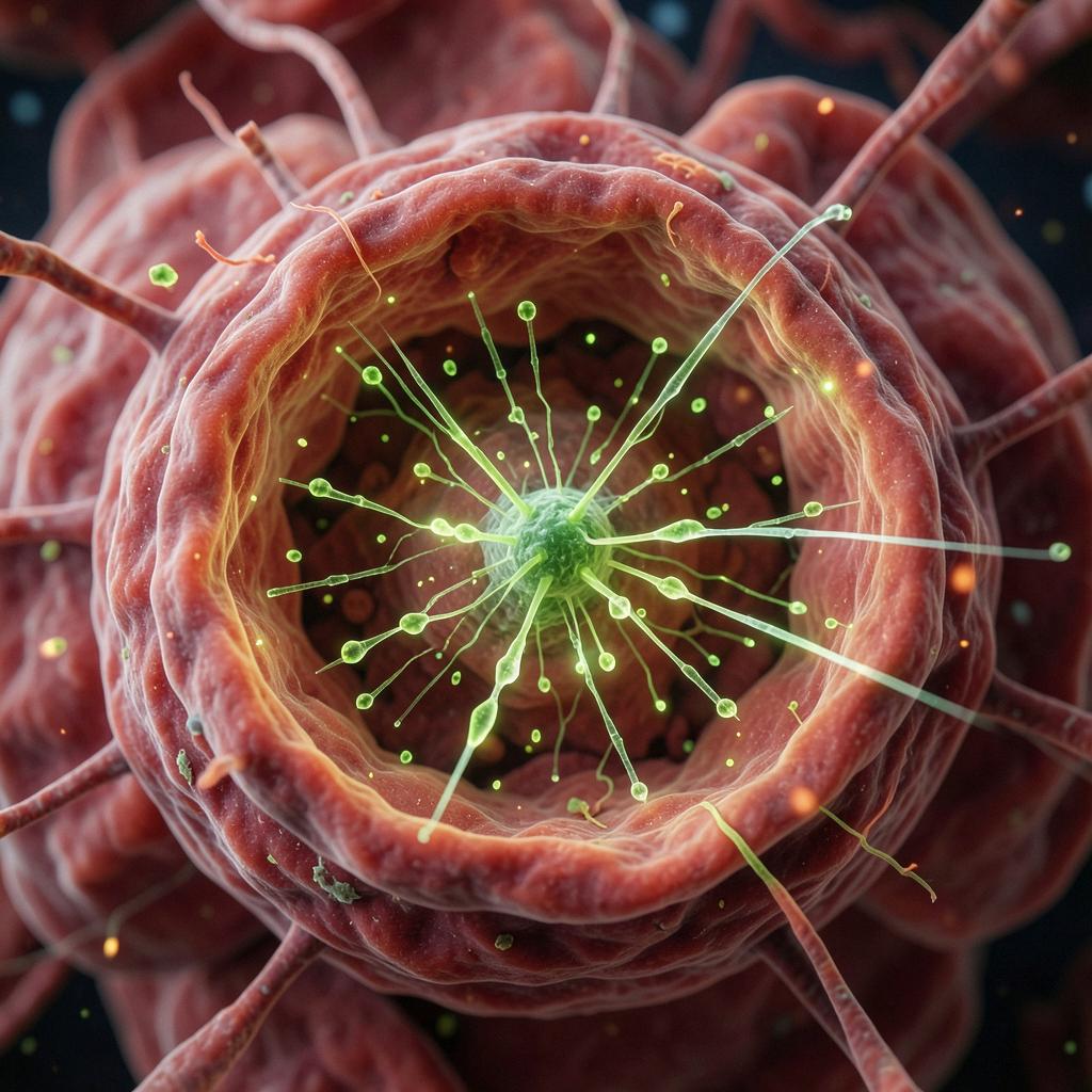
 Science Movie
 Discussion