Ask questions across claims linked to experiments, exact results, limitations, and sources.Know what falsifiable science actually supports before you trust the answer.
Press Enter ↵ to search evidence
Explore by Goal
"In biology, nothing is clear, everything is too complicated, everything is a mess, and just when you think you understand something, you peel off a layer and find deeper complications beneath. Nature is anything but simple."
- Richard Preston
Quick Explanation
Copied
The paper reports a PVA/Gel-A full-IPN nanofibrous hydrogel loaded with bromothymol blue that shows +216% modulus, <10% dye leaching with PDADMAC, <1 min color response across pH 4–9, and acceptable KeratinoSens cytocompatibility, supporting its promise as a smart wound dressing pending in vivo validation (doi:10.1039/d5tb01305d).
Long Explanation
What the Study Demonstrates
The work fabricates electrospun PVA nanofibers converted into a full interpenetrating network (IPN) with a phosphine oxide-based Gel-A via citric acid crosslinking and 130 °C post-treatment. The 1:1 PVA/Gel-A composition improved modulus (+216%), tensile strength (+154.5%), and elongation (+58%) versus pure PVA fibers (doi:10.1039/d5tb01305d).
Sensing Performance
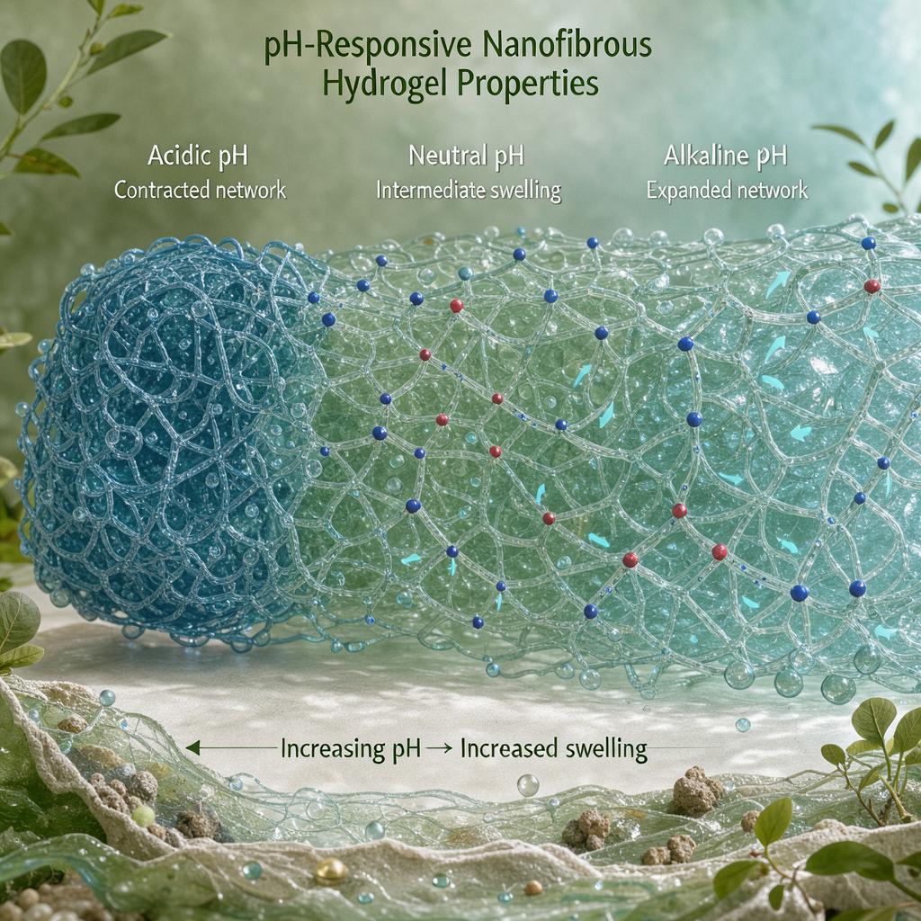
Bromothymol blue (2.5 mg/mL) immobilized with the cationic complexing agent PDADMAC (6 mg/mL) reduced dye leaching to <10%. BTB-loaded fibers (440 ± 130 nm diameter; unloaded fibers 675 ± 136 nm) shifted UV-Vis λmax from 434 nm at pH 5 to 616 nm at pH 8, with visible color changes in under 1 minute; Kubelka–Munk K/S peaks shifted from 440 nm (no PDADMAC) to 420/620 nm with PDADMAC (doi:10.1039/d5tb01305d).
Cytocompatibility and Context
KeratinoSens assays (three independent extractions, five independent experiments per assay) showed >70% viability and sensitization fold-induction <1.5, meeting OECD 442D thresholds. BET analysis showed the composite shifted from mesoporous (PVA, 10.3 m²/g) to microporous (2.52 m²/g), and zeta potential indicated dual charging with an isoelectric point near pH 4 (doi:10.1039/d5tb01305d).
Critical Caveats
Claims rest entirely on in vitro data: no wound-model or in vivo infection correlation was performed, and the paper infers infection monitoring from pH proxy rather than direct biomarkers (e.g., bacterial load). PDADMAC in vivo safety, long-term BTB stability in exudate, and scalability remain unaddressed. Only a single 1:1 composition was characterized in depth; Gel-A brittleness was acknowledged. These gaps temper the smart-dressing translation claim despite strong material-level evidence (doi:10.1039/d5tb01305d).
Feedback:
Updated: November 18, 2026
BGPT Paper Review
Study Novelty
80%
Combining a phosphine oxide-based Gel-A into a full-IPN with PVA for leach-resistant colorimetric wound pH sensing is a materially novel composition, though pH-indicator dressings themselves are established.
Scientific Quality
80%
Well-characterized (SEM, ICP-OES, BET, tensile, zeta, UV-Vis, OECD 442D assays) with clear replicates; but claims exceed the in vitro scope, and no in vivo wound-model data are provided.
Study Generality
70%
Platform approach could generalize to other dyes/indicators, but results are specific to one PVA/Gel-A ratio and BTB chemistry.
Study Usefulness
80%
Directly relevant to smart wound dressing development; mechanical and dye-retention gains are practically meaningful for clinical translation, contingent on in vivo confirmation.
Study Reproducibility
70%
Detailed methods, instrument models, and SI data support replication; however, gel synthesis details and batch variability reporting are not fully transparent.
Explanatory Depth
80%
Mechanistic links between IPN structure, dual-charging zeta behavior, ionic dye interactions, and colorimetric response are well developed via Kubelka-Munk analysis.
We'll email you the results when your analysis is finished.
Hypothesis Graveyard
BTB leaching is dominated by hydrophilic diffusion through swollen PVA alone; rejected because adding PDADMAC reduced leaching to <10% without major swelling changes, implicating ionic complexation as the dominant retention mechanism.
Fiber thinning after dye loading (675 to 440 nm) is electrospinning variability; more plausible that dye/PDADMAC plasticization and jet conductivity changes altered fiber formation, warranting conductivity controls.

 Explore by Goal
 Quick Explanation
 Long Explanation
 Top Data Sources
 Ask a Follow-Up
 Key Insight
 Keep Exploring
 Hypothesis Graveyard
 Potential Experiments
 Discussion
