Analyze how experimental results and claims are presented, scoped, and limited in a paper.Know what the science actually supports before you trust the answer.
Press Enter ↵ to analyze
Explore by Goal
"Biology sees right and wrong as the same color in different light."
- Delia Owens
Quick Answer
Copied
This 2005 JBC study used retroviral shRNA to show that ARD1 knockdown (>80% mRNA reduction) does not stabilize HIF-1α or induce hypoxia-responsive genes, contradicting the prior ARD1-HIF acetylation model, and instead identifies ARD1 as required for maintaining cell proliferation in HepG2 cells.
Long Answer
Core Finding: ARD1 Is Not Required for HIF Suppression
Fisher et al. (2005) directly tested the hypothesis that ARD1, proposed as a HIF-1α acetylase regulating its stability, is required for hypoxia response. Using two independent retroviral shRNA hairpins in HepG2 cells, they achieved 85% (hairpin 1.3) and 35% (hairpin 1.6) ARD1 mRNA knockdown. Critically, neither hairpin altered HIF-1α protein levels under normoxia or prevented HIF-1α stabilization under hypoxia. Gene chip analysis of seven sample conditions (triplicates, n=21) confirmed that erythropoietin, VEGF, EGLN1, and EGLN3 were induced by hypoxia but showed no change under ARD1 knockdown alone or combined with hypoxia beyond additive effects
This result contradicts the prior model by Jeong et al. (2002) claiming ARD1 acetylates HIF-1α at Lys532 to promote pVHL-mediated degradation .
ARD1 Regulates Proliferation and Metabolism
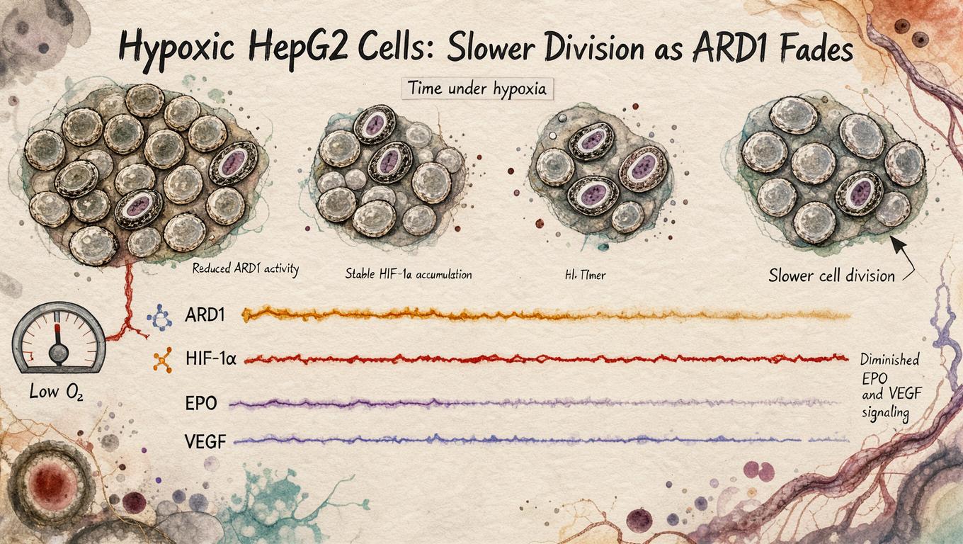
Gene chip analysis revealed that the 10 most downregulated genes in ARD1-knockdown cells (hairpin 1.3) included proliferation promoters (TPD52, BECN1) and metabolic enzymes (PGM3, GLRX), while anti-proliferative genes (BIK) and extracellular matrix genes (COL2A1, FBN1, LGALS7) were upregulated. ARD1 overexpression (4.5-fold) induced cell division control protein 2 (CDC2). CFSE dilution assays confirmed dramatic proliferation slowing by day 7, with mean fluorescence intensity rising from 11.92 (control Kk) to 24.77 (1.6 RNAi) and 29.54 (1.3 RNAi), confirming ARD1's pro-proliferative role . This is consistent with later findings that ARD1 acetylates PGK1 to activate Beclin1-dependent autophagy under hypoxia .
Decreased EPO/VEGF Is a Proliferation Artifact
Although ELISA showed lower erythropoietin and VEGF protein in ARD1-knockdown supernatants, mRNA levels (via qRT-PCR and gene chip) were unchanged. The authors attribute reduced protein output to slower cell proliferation rather than HIF-dependent transcriptional changes — a logical but underpowered interpretive step lacking direct normalization to cell number .
Green = upregulated by hypoxia; Red = downregulated. Data from Table I of the paper .
Critical Limitations
Single cell line focus. HepG2 was chosen for retroviral transduction efficiency and EPO production, but the authors acknowledge Hep3B produces ~10-fold more EPO and that cell-type-specific hypoxia responses differ qualitatively across lines. Generalization to in vivo tissue hypoxia remains untested. Compensation not ruled out in vivo. No compensatory upregulation of ARD2 was detected in HepG2 (extremely low baseline), but the study acknowledges that ARD2 is expressed in K562 cells, leaving open cell-context-dependent redundancy . Knockdown ≠ knockout. Residual ARD1 (15% with hairpin 1.3) could suffice for HIF regulation; the dose-response relationship between ARD1 level and HIF stability was not established.
What would change the conclusion: Evidence that ARD1 acetylation-deficient HIF-1α mutants (e.g., K532R) are stabilized under normoxia independent of ARD1 protein levels, or that complete ARD1 knockout (not partial knockdown) stabilizes HIF-1α.
Context in ARD1 Biology
Later work confirmed ARD1's role in proliferation across systems: hARD1 overexpression enhanced MCF-7 breast cancer cell proliferation (3.48-fold vs 2.35-fold control) and hARD1 was elevated in 86% of breast cancer tissues . In Drosophila, Ard1 null alleles are embryonic-lethal and ovary-specific knockdown causes pleiotropic oogenesis defects including desynchronized cystocyte divisions, reinforcing a conserved pro-proliferative function .
Feedback:
Updated: November 03, 2026
BGPT Paper Review
Study Novelty
60%
Challenges the established ARD1-HIF acetylation model using loss-of-function genetics rather than overexpression, a meaningful but incremental advance in 2005.
Scientific Quality
70%
Well-controlled with two independent shRNAs, gene chip validation, and CFSE proliferation assay. Weaknesses include single-cell-line focus, partial knockdown not full knockout, and lack of HIF acetylation site-specific analysis.
Study Generality
50%
Findings limited to HepG2/293T cell lines in vitro; cell-type-specific hypoxia responses reduce generalizability to in vivo hypoxia biology.
Study Usefulness
70%
Redirected the field from ARD1-as-HIF-regulator toward ARD1-in-proliferation, anticipating later discoveries in NatA biology, autophagy, and cancer metabolism.
Study Reproducibility
70%
Detailed methods with primer sequences, vector constructs, and statistical analysis reported; gene chip data lack accession numbers.
Day 7 values are observed; intermediate timepoints interpolated for visualization only. Higher CFSE = fewer divisions. Assumes exponential CFSE dilution and no dye transfer between cells .
Generated scientific data; not direct experimental measurements.
Get emailed when your analysis is done!
We'll email you the results when your analysis is finished.
Hypothesis Graveyard
ARD1 is a physiologically relevant HIF-1α acetylase controlling its stability via Lys532 — rejected by this paper's independent shRNA approach showing no HIF stabilization upon >80% ARD1 depletion in two cell lines, and by lack of replication of the Jeong et al. overexpression-based result.

 Explore by Goal
 Quick Answer
 Long Answer
 Top Data Sources
 Ask a Follow-Up
 Key Insight
 Keep Exploring
 DataGen
 Hypothesis Graveyard
 Discussion
