Check whether claims match experiments, exact reported results, and stated limitations.Know what the science actually supports before you trust the answer.
Press Enter ↵ to review paper
Explore by Goal
"I think it's much more interesting to live not knowing than to have answers which might be wrong."
- Richard Feynman
Quick Explanation
Copied
This 1976 electron-microscopy study of Hyalophora cecropia prothoracic glands showed that the first 3–4 days of cytoplasmic changes after cold release occur identically in brainless and methylxanthine-inhibited pupae, while full secretory differentiation (RER expansion, vesicle release, basement-membrane breakdown) requires brain hormone.
Long Explanation
What the paper showed
McDaniel, Johnson, Saum and Berry compared prothoracic gland ultrastructure in four groups of diapausing Hyalophora cecropia pupae: untreated controls (pharate adults), debrained dauer pupae, and pupae arrested with aminophylline (a methylxanthine) either before or after chilling. After transfer from 4°C to 22°C, all groups showed the same early sequence for 3–4 days: large membrane-unbounded vacuoles disappeared, free ribosomes and microtubules increased, glycogen rose then vanished, and small RER cisternae, Golgi bodies, ring nucleoli and annulate lamellae appeared. Only in brains-intact pharate adults did day 4–5 bring secretory differentiation: extensive RER with flocculent material, cytoplasmic vesicles released between microvillae, basement-membrane disruption and microvillar resorption. Dauer and aminophylline-arrested glands remained frozen at the day-4 state for over 2–3 months
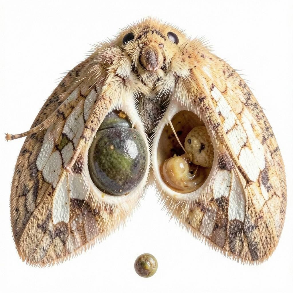
Qualitative schematic reconstructed from the paper's text description of ultrastructural milestones; the authors report no quantitative morphometry.
Critical assessment
Strengths. The design cleverly separates temperature-driven cellular responses from brain-hormone-dependent secretion by using dauer pupae and drug-arrested animals as negative controls—prior ultrastructural studies had only examined intact animals, which could not distinguish these two causes . The single dauer pupa that spontaneously initiated development showed the brain-intact phenotype (vesicles, RER, disrupted basement membrane), a natural internal control supporting the interpretation.
Limitations and blind spots. Sampling was at least two animals per group per day; no morphometric quantification, no statistics, and consistent cell-to-cell lagging within glands are acknowledged. The aminophylline mechanism (proposed blockade of brain-hormone release) is inferential—later work complicates this, since aminophylline stimulates ecdysone secretion from Manduca glands in vitro via cAMP/phosphodiesterase effects , making direct inhibition of gland output an alternative explanation the paper under-explores. No direct assay of ecdysone or PTTH titers was performed; the mechanism rests entirely on morphology. Species generality is limited, though Manduca work later confirmed multi-factor gland regulation (PTTH forms, hemolymph factor, juvenile hormone competence) .
Confidence note. The central descriptive finding (arrest at the day-4 ultrastructural state without brain hormone) is well-supported by the reported serial sampling; the causal attribution to brain hormone is plausible but mechanism-level, unmeasured, and would be falsified if ecdysone or PTTH assays showed secretion resuming in arrested glands.
Feedback:
Updated: October 26, 2026
BGPT Paper Review
Study Novelty
60%
First study to dissect brain-hormone-dependent vs temperature-dependent ultrastructural changes in prothoracic glands using debrained and drug-arrested controls; significant for 1976 but now standard insect endocrinology.
Scientific Quality
70%
Careful serial EM sampling with appropriate negative controls and an internal positive control (spontaneously developed dauer pupa); weakened by tiny sample sizes (≥2 animals/group/day), no quantification, no hormone titers, and reliance on morphological inference.
Study Generality
50%
Single species (H. cecropia), single tissue, specific diapause paradigm; the conceptual point (environmental vs hormonal control of secretory differentiation) generalizes, but the descriptive data are niche.
Study Usefulness
60%
Established that brain hormone is required for the late secretory phase of gland activation, informing later PTTH/cAMP/hemolymph-factor work on ecdysteroidogenesis regulation.
Study Reproducibility
60%
Methods (fixation, staging, temperature/photoperiod regimens, drug doses) are clearly described and feasible, but qualitative EM scoring without morphometric criteria makes exact replication of stage calls operator-dependent.
Explanatory Depth
60%
Provides a clear two-phase mechanistic model (temperature response vs hormone-dependent secretion) but lacks molecular mechanisms, hormone measurements, or direct tests of the aminophylline target.

 Explore by Goal
 Quick Explanation
 Long Explanation
 Top Data Sources
 Ask a Follow-Up
 Key Insight
 Keep Exploring
 Hypothesis Graveyard
 Potential Experiments
 Discussion
