Analyze how experimental results and claims are presented, scoped, and limited in a paper.Know what the science actually supports before you trust the answer.
Press Enter ↵ to analyze
Explore by Goal
"The finding of the double helix thus brought us not only joy but great relief. It was unbelievably interesting and immediately allowed us to make a serious proposal for the mechanism of gene duplication."
- James Watson
Quick Explanation
Copied
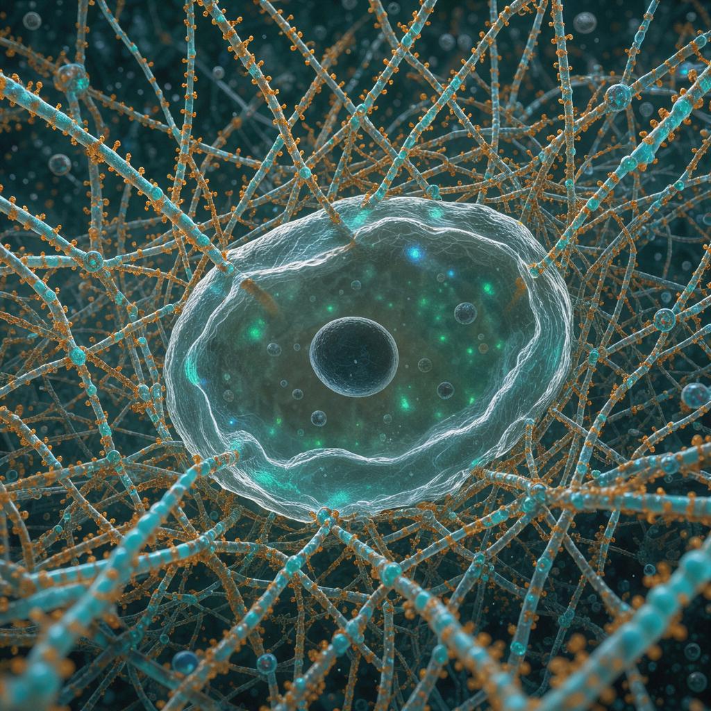
This 2005 Acta Biomaterialia paper by Beniash, Hartgerink, Storrie, Stendahl, and Stupp established that peptide amphiphiles self-assemble into ~5–6 nm nanofiber hydrogels at physiological pH via polyvalent metal ions, entrapping MC3T3-E1 cells that survive ≥3 weeks, proliferate, and endocytose nanofibers, with the EQS-terminated variant proving toxic — a foundational but cell-line-limited and largely observational study.
Long Explanation
Core Findings (Reported Evidence)
The paper demonstrates metal-ion-triggered self-assembly of peptide amphiphiles (PAs) into 5–6 nm cylindrical nanofibers at physiological pH, with TEM showing uranyl-acetate-stained charged peripheries and unstained hydrophobic cores, and FTIR indicating parallel β-sheet peptide packing (amide I at 1630–1640 cm⁻¹). Rheology confirmed gel character (G′ > G″) for polyvalent-ion gels, while monovalent ions yielded viscous liquids — except IKVAV-containing PAs 7 and 8, which gelled with 200 mM KCl, attributed to hydrophobic side-chain associations in the IKVAV motif .
Cell entrapment was sequence-dependent: KGE-terminated PA 3 gels supported survival, ~6-fold proliferation over 8 days to a ~60,000 cells/mL plateau, spindle-shape adoption, and aggregation; EQS-terminated PA 5 gels killed all cells by day 20. TEM showed cells endocytosing nanofibers into lysosome-like compartments, and positive R(DG,DL) (lactate + glucose mass change) suggested cells metabolized PA molecules as nutrients .
Note: only the fold-change and plateau density are reported; intermediate time points were not provided, so the curve is a two-point rendering of stated values, not a full trajectory.
Critical Assessment
Strengths: Multi-technique convergence (rheology, TEM, FTIR, UV–Vis Cu-complexation, metabolic assays) and honest reporting of a toxic variant (PA 5), a negative result rarely disclosed. Gel stability (pH 4–11, 100 °C, 14 days in 10× water) is a genuine advance over Ca-alginate. The metal-ion gelation mechanism solves the pH-instability problem of earlier pH-triggered PA assembly, and the cell-entrapment concept proved foundational — PA systems remain central to 3D microenvironment engineering in later reviews .
Blind spots: Single cell line (MC3T3-E1) and cell type; toxicity mechanism of PA 5 unexplained (authors only speculate on cancer-therapy use); no quantified elasticity values per ion despite rheology claims; metabolic inference from R(DG,DL) is indirect — amino acid metabolism into lactate could inflate lactate independent of PA catabolism, and no isotope tracing was performed; KGE assumed integrin-inert based on a single literature report, never tested with blocking antibodies. The 200 mM–6 M ion-screening lacks cytotoxicity corroboration for non-gelating conditions. Funding was DOE/NSF with no declared conflicts — appropriate disclosure for a non-clinical materials study.
Uncertainty: The "cells utilize PA molecules in metabolic pathways" claim is an author interpretation; direct evidence (radiolabeling) is absent. Whether endocytosed nanofibers are degraded or accumulate over weeks longer than 20 days is unknown.
What Would Change the Conclusion
Isotope-labeled PA tracing failing to show PA-derived carbon in lactate would falsify the nutrient-utilization claim; broad-spectrum cell-line testing showing PA 3 also kills primary cells would undermine the tissue-engineering premise. Conversely, follow-up work on PA hydrogels in regenerative medicine has broadly validated biocompatibility, supporting the core findings .
Explore Further
Author Reviews
Feedback:
Updated: October 31, 2026
BGPT Paper Review
Study Novelty
80%
First demonstration of metal-ion-triggered PA nanofiber gelation at physiological pH for live-cell entrapment with evidence of cellular endocytosis and metabolic utilization of the scaffold — a novel mechanism within the authors' established PA platform (prior work used pH triggers).
Scientific Quality
80%
Multi-method characterization (rheology, TEM, FTIR, UV-Vis, metabolic assays) with appropriate controls (monovalent vs polyvalent ions, ion-depleted media, gel-free controls) and candid disclosure of the toxic PA-5 variant. Weaknesses: single cell line, indirect metabolic inference, no quantified moduli reported, unproven KGE-integrin-inertness assumption.
Study Generality
70%
Establishes a general self-assembly mechanism applicable to multiple PA sequences and 8 metal ions, but cell findings rest on one immortalized osteoblast-like line, limiting generalization to other tissues.
Study Usefulness
80%
The physiological-pH metal-ion trigger enabled injectable/in-situ gelling PA matrices, later foundational to neural, bone, and other tissue-engineering applications; the sequence-dependent cytotoxicity finding also informs PA library screening.
Study Reproducibility
70%
Synthesis, assembly, rheometry, microscopy, and cell protocols are described in enough detail for replication; however, exact moduli values, replicate counts for cell experiments, and raw metabolic data are not fully tabulated.
Explanatory Depth
70%
Mechanism is mechanistically reasoned (electrostatic bridging of acidic residues by polyvalent ions, β-sheet hydrogen bonding, hydrophobic core), supported by structure–function comparisons across 8 PAs; but metal-ion coordination geometry and the PA-5 toxicity mechanism remain unresolved.
We'll email you the results when your analysis is finished.
Hypothesis Graveyard
KGE activity as a cryptic integrin ligand: unlikely because RGD→RGE substitution reduces fibronectin binding >100-fold per cited literature, and authors assumed inertness; direct blocking-antibody tests were nonetheless never performed.
Gel toxicity from residual free metal ions: unlikely since gel-free control cultures in the same medium showed normal R(DG,DL), though ion scavenging controls were not explicitly run.

 Explore by Goal
 Quick Explanation
 Long Explanation
 Top Data Sources
 Ask a Follow-Up
 Key Insight
 Keep Exploring
 Hypothesis Graveyard
 Potential Experiments
 Discussion
