Quickly verify claims by accessing the underlying experimental data and figures.
Press Enter ↵ to solve
Fuel Your Discoveries
"The universe is not only queerer than we suppose, but queerer than we can suppose."
- J.B.S. Haldane
Quick Answer
Copied
Core finding
In STZ-diabetic male C57BL/6J mice, Nlrp3 knockout reduced renal inflammation, oxidative stress, and fibrosis markers, with decreased IL-1β/IL-18 axis readouts, TGF-β1/CTGF expression and Smad3 activation, and lower TXNIP/Nox4/superoxide and urinary 8-OHdG.
Mechanistic claim is supported by genetic (NLRP3 KO/shRNA) + antioxidant (tempol) + cytokine (IL-1β exposure) perturbations in vivo and HK-2 cells, but causal direction beyond the tested axis (e.g., pyroptosis vs noncanonical NLRP3 functions; cell-type specificity; complete IL-1β dependence) is not fully established in the provided text.
Long Answer
Paper review (evidence-based, skeptical): NLRP3 deficiency ameliorates renal inflammation and fibrosis in diabetic mice
DOI: 10.1016/j.mce.2018.08.002 • Publication date metadata in provided record: December 01, 2018 (journal listing) • Focus: STZ-diabetic mouse kidney + HK-2 high-glucose model + TXNIP/Nox4/ROS axis.
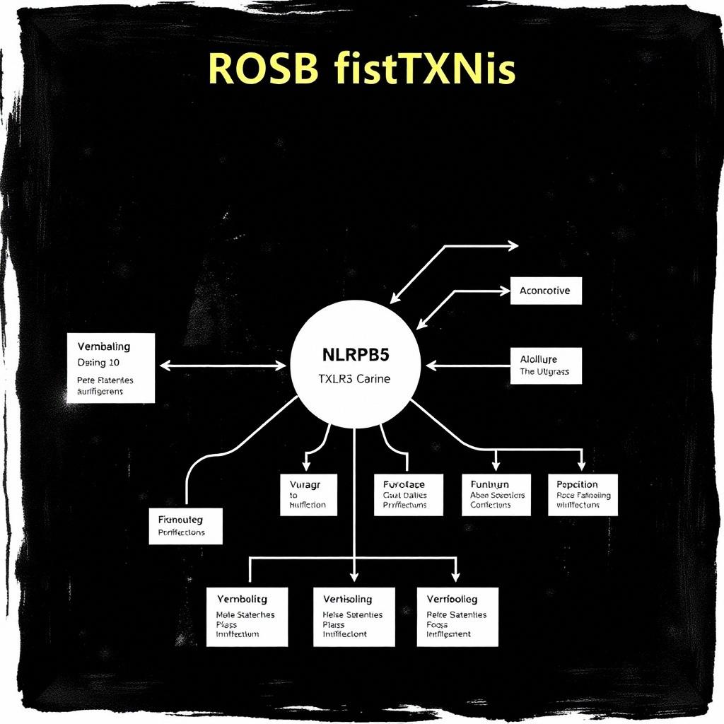
VISUAL 1 — Claimed mechanistic axis (as presented in the paper)
The study proposes that NLRP3 activity contributes to diabetic kidney inflammation and fibrosis at least partly via oxidative stress, connecting TXNIP → Nox4 → superoxide/ROS, and that IL-1β can further drive ROS/TXNIP/Nox4 in tubular cells.
VISUAL 2 — Directional renal benefit of NLRP3 KO in the paper
Qualitative summary (because the provided full-text extract does not include numeric effect sizes for every endpoint). Blue = decreased diabetes effect with KO; gray = “not changed by KO” (diabetes glucose not affected).
EXPLAIN 1 — What the authors actually did (and what that supports)
In vivo model: Male C57BL/6J STZ-induced diabetes; harvested at 24 weeks; NLRP3-/- mice compared to WT littermates; diabetes severity (blood glucose) reported as not changed by NLRP3 KO.
Kidney readouts: kidney morphologic scoring (PAS/Masson), TEM for GBM thickness/foot process effacement, Western blot and IHC/IF for ECM (fibronectin, collagen I/IV), inflammasome/proinflammatory molecules (caspase-1 p10, cleaved IL-1β, IL-18; MCP-1; macrophage marker F4/80), profibrotic TGF-β1/CTGF and Smad3 phosphorylation, and oxidative stress markers (TXNIP, Nox4, superoxide; urinary 8-OHdG).
In vitro cell model: HK-2 proximal tubular epithelial cells under high glucose ± osmotic control (mannitol), with NLRP3 shRNA or tempol, and IL-1β stimulation; ROS assessed with intracellular DCFDA-like probe by flow cytometry and mitochondrial ROS via MitoSOX; superoxide via lucigenin assay.
EXPLAIN 2 — Evidence strength for the paper’s main causal chain (what is supported vs not fully proven)
“NLRP3 KO improves diabetic kidney outcomes” — Supported by multiple independent endpoint families in the same experiment (function proxies, histology, ECM markers, inflammasome/inflammatory markers, TGF-β/Smad profibrotic markers, oxidative markers). The paper also reports blood glucose is not altered by NLRP3 KO, which partially reduces (but does not eliminate) a metabolic confounding explanation.
“NLRP3 affects oxidative stress via TXNIP/Nox4” — Supported by concordant in vivo downshift of TXNIP and Nox4 with KO and decreased superoxide/8-OHdG, plus in vitro prevention of high-glucose-induced TXNIP and Nox4 expression and ROS generation by NLRP3 shRNA and by the antioxidant tempol. However, the paper does not (in the provided extract) include a “rescue” experiment proving that restoring TXNIP/Nox4 reinstates the phenotype specifically downstream of NLRP3.
“Inflammasome/IL-1β participates upstream and can drive ROS/TXNIP/Nox4” — The in vitro IL-1β stimulation portion shows IL-1β can induce TXNIP and Nox4 and increase ROS in HK-2 cells, which supports plausibility that IL-1β can connect inflammation to oxidative signaling. What is less established in the extract is whether IL-1β blockade (e.g., IL-1 receptor antagonism or IL-1β neutralization) is sufficient to mimic NLRP3 deficiency for the oxidative/fibrotic endpoints in vivo.
VISUAL 4 — Context: how this paper fits broader NLRP3–kidney inflammation/fibrosis knowledge
The paper aligns with broader claims that NLRP3 contributes to renal inflammation/fibrosis in CKD contexts, while also emphasizing a potentially inflammasome-linked and oxidative stress–linked pathway via TXNIP.
The TXNIP/NLRP3/IL-1β oxidative axis is consistent with related diabetic nephropathy work highlighting mitochondrial ROS → TXNIP → NLRP3/IL-1β signaling.
SKEPTICAL CRITIQUE — limitations, blindspots, and what would most disprove the main claims
Single disease model + sex: The study uses STZ-induced diabetic mice and only reports male animals (as provided in methods section). Generalization to other DN etiologies and to females is uncertain.
Cell-type specificity of NLRP3 action: NLRP3 KO is systemic. The extract does not include cell-type–specific knockouts to demonstrate whether renal tubular cells vs immune infiltrates vs fibroblasts are the dominant causal node.
Mechanistic “necessity” gaps: The study shows NLRP3 deficiency reduces TXNIP/Nox4/ROS and that IL-1β can induce TXNIP/Nox4/ROS in HK-2. But the extract does not show (i) TXNIP overexpression rescue downstream of NLRP3 KO, or (ii) IL-1β blockade proving IL-1β is necessary for the oxidative/fibrotic phenotype.
ROS measurement specificity: ROS assays (CM-DCHF-DA, MitoSOX, lucigenin chemiluminescence) are sensitive to probe chemistry and experimental conditions; the extract does not describe orthogonal validation (e.g., multiple mitochondrial-specific readouts beyond MitoSOX, or controls for probe oxidation artifacts). This is a common interpretability issue for ROS studies (not a claim that the paper is wrong; just an uncertainty to consider).
Most discriminating falsification tests (conceptual):
Show that preventing TXNIP/Nox4/ROS (via TXNIP-independent approaches) does not replicate the full NLRP3 KO phenotype, implying additional NLRP3 pathways contribute to fibrosis/inflammation.
Show that reintroducing TXNIP (or constitutively active Nox4 signaling) into NLRP3 KO contexts restores ROS/inflammation/fibrosis endpoints.
Show that IL-1β axis blockade removes NLRP3 KO benefits—or conversely that IL-1β blockade fails to fully remove benefits—clarifying whether IL-1β is a key mediator vs a parallel marker.
VISUAL 5 — Evidence triage by claim level (paper-internal)
Levels: Association (correlation-like observation), Intervention support (genetic/chemical perturbation affects outcomes), Mechanism necessity (not fully established from provided extract).
Actionable takeaways for the reader
If you’re mapping inflammation→oxidative stress→fibrosis in DN, this paper provides an experimentally linked axis between NLRP3 and TXNIP/Nox4/ROS readouts in kidney tissue and HK-2 cells.
For mechanistic rigor, the “next step” experiment space is clear: test necessity/rescue for TXNIP and IL-1β, and use cell-type–specific NLRP3 models to prevent attribution ambiguity. (These are not claimed by the paper; they are what would most improve interpretability.)
Author reviews on BGPT
Feedback:
Updated: March 29, 2026
BGPT Paper Review
Study Novelty
70%
The novelty is the specific integration of NLRP3 deficiency with a kidney oxidative-stress axis centered on TXNIP/Nox4/ROS and coupling IL-1β’s downstream effects in HK-2 cells, rather than just reporting NLRP3→inflammation or NLRP3→fibrosis in isolation. This is incremental relative to the broader NLRP3–DN literature, but the mechanistic triangulation is relatively specific to this paper’s axis.
Scientific Quality
70%
Strengths: multi-level phenotype (function proxies, histology/ECM, inflammasome/inflammatory markers, TGF-β/Smad signaling, oxidative stress) and multi-perturbation design (global KO; shRNA; tempol; IL-1β exposure). Main quality limitations (from the provided extract) are gaps in necessity/rescue and cell-type specificity, and reliance on probe-based ROS assays without orthogonal confirmation described in the extract.
Study Generality
60%
Generality is moderate: results come from STZ-diabetic mice and HK-2 cells, so extrapolation to other DN etiologies and human disease heterogeneity is uncertain. The mechanistic axis is plausible within CKD inflammatory/oxidative frameworks, but the paper does not establish universal causality across models.
Study Usefulness
70%
Usefulness is solid for mechanistic hypothesis generation and for guiding what assays/markers to include when studying NLRP3/TXNIP/ROS in DN. However, translational usefulness is limited because causal necessity is incomplete and the extract provides no clinical validation.
Study Reproducibility
60%
Reproducibility is moderate: methods are reasonably described (STZ protocol, assays, KO model, HK-2 culture conditions, tempol and shRNA usage, ROS/superoxide measurement approaches, ImageJ quantification). Reproducibility uncertainty remains because the extract does not provide full details such as exact blot quantification raw values, randomization/blinding procedures for all endpoints, and complete primer sequences/replicates for every assay.
Explanatory Depth
70%
Depth is fairly strong for a mechanistic preclinical study: it ties NLRP3 perturbation to an oxidative stress axis (TXNIP/Nox4/ROS) and links IL-1β to downstream ROS/TXNIP/Nox4 induction in tubular cells. Depth is limited by incomplete causal necessity/rescue demonstrations and lack of cell-type–specific genetic dissection in vivo within the extract.
We'll email you the results when your analysis is finished.
Hypothesis Graveyard
“NLRP3 KO improves DN mainly by lowering systemic blood glucose.” The paper reports blood glucose is not affected by NLRP3 KO, making this explanation less consistent with the presented data.
“The IL-1β–TXNIP/Nox4 relationship is an artifact with no functional downstream relevance.” This is disfavored by the paper’s IL-1β induction experiments in HK-2 cells combined with concurrent in vivo reduction in cleaved IL-1β/IL-18 and downstream oxidative/profibrotic markers with NLRP3 KO.