Analyze how experimental results and claims are presented, scoped, and limited in a paper.Know what the science actually supports before you trust the answer.
Press Enter ↵ to analyze
Explore by Goal
"Be less curious about people and more curious about ideas."
- Marie Curie
Quick Explanation
Copied
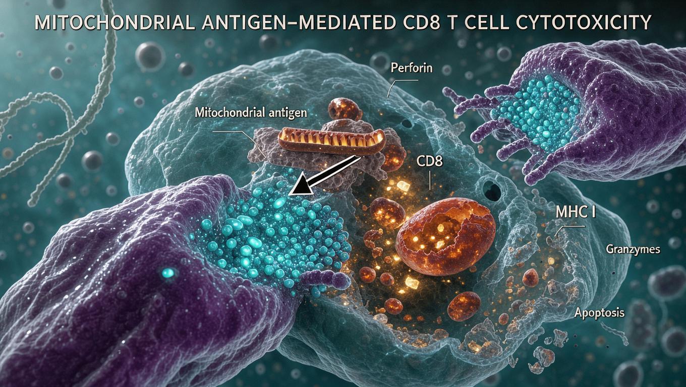
This bioRxiv preprint (10.1101/2024.02.26.582098) provides the most direct causal test to date that mitochondrial antigen-specific CD8⁺ T cells alone are sufficient to infiltrate the brain, persist as functional memory cells, and drive selective nigrostriatal dopamine neuron loss with levodopa-reversible motor deficits in mice — but it relies on an artificial adoptive-transfer model with pertussis toxin and pre-activated T cells, and human generalization, mechanism of killing, and glial involvement remain unresolved.
Long Explanation
Core Finding and Evidence Chain
The study adoptively transferred 5×10⁶ in vitro-activated OGDH-specific CD8⁺ T cells (Mito-T, from 2C TCR transgenic donors) into WT and Pink1⁻/⁻ mice, with OT-I-derived ovalbumin-specific cells (OVA-T) as controls; pertussis toxin (20 µg/kg) was given to open the blood-brain barrier. Key results: (1) Mito-T cells entered the brain and persisted at least 40 days, with higher absolute counts in Pink1⁻/⁻ recipients; (2) recipients showed reduced open-field movement, rotarod deficits, and pole-test impairment that was fully reversed by L-DOPA/benserazide, the hallmark dopaminergic fingerprint; (3) stereology revealed significant SNc TH⁺ neuron loss with sparing of VTA DA neurons, SERT⁺ terminals, and NeuN⁺ cells — selective dopaminergic vulnerability; (4) infiltrating cells acquired CD44⁺CD127⁺CXCR3⁺CXCR6⁺ memory/Tc1 phenotypes retaining IFN-γ, TNF-α, and granzyme B production. Critically, degeneration occurred in both genotypes, suggesting PINK1's protective role may lie in peripheral priming restriction, not neuronal-intrinsic survival .
Scientific Context and Plausibility
The work builds directly on Matheoud et al., who showed PINK1/Parkin repress mitochondrial antigen presentation on MHC-I and that intestinal infection in Pink1⁻/⁻ mice triggers parkinsonism — but that prior work did not isolate T cells as sufficient . The mechanistic plausibility of neurons as MHC-I-dependent CD8⁺ T cell targets is independently established: Borna virus-infected neurons upregulate MHC-I (~30.6% of neurons vs ~0.56% baseline), and MHC-I blockade restores CD8⁺ T cell motility and reduces neuronal apoptosis .
Critical Limitations and Blindspots
(1) Artificial sufficiency: pre-activated monoclonal TCR cells plus PTx-mediated BBB disruption does not model spontaneous disease onset; the authors acknowledge this reductionism but argue it cleanly isolates one causal question. (2) Mechanism of kill is unresolved: whether DA neurons present OGDH peptide themselves or glia mediate indirect toxicity was not tested; MHC-I expression on DA neurons in vivo was not quantified. (3) No correlation data: the paper does not report T cell counts vs neuron loss per mouse, weakening dose-response inference. (4) Incomplete glia assessment: only Iba1/GFAP markers at 30 days were examined — normalizing at a single timepoint cannot exclude earlier functional glial activation. (5) Translation risk: OGDH T cell frequency in human PD blood is unestablished here; murine results pooled sexes with unstated per-group n. (6) Preprint status: not yet peer-reviewed at extraction; conclusions framed as "most direct support to date" warrant cautious reading pending review .
What Would Falsify or Extend the Model
Falsification routes: blocking MHC-I in vivo or depleting granzyme/perforin should abolish degeneration; endogenous (non-transfer) TCR sequencing in aged WT mice should show OGDH-reactive clonotypes; human validation would require OGDH-specific TCR frequency correlating with PD progression. If glial MitAP proves dominant, the direct-killing framing shifts to a neuroinflammation-amplification model. The surprising WT degeneration, if replicated, would reframe PINK1 as a peripheral tolerance gatekeeper — a genuinely provocative, testable repositioning of the field's dominant mitophagy-centric narrative.
Explore Further
Author Reviews
Feedback:
Updated: November 02, 2026
BGPT Paper Review
Study Novelty
90%
First direct sufficiency test that brain-infiltrating mitochondrial antigen-specific CD8⁺ T cells alone cause parkinsonism; prior work only correlated T cell expansion with infection-triggered symptoms.
Scientific Quality
80%
Rigorous controls (OVA-T, PTx-only), blinded behavior, unbiased stereology, multiple independent replicates. Limits: preprint status, artificial transfer with PTx, unresolved killing mechanism, no T-cell-count/neuron-loss correlation, pooled sexes with unstated per-group n.
Study Generality
60%
Mouse-specific adoptive-transfer paradigm; broad implications for PD autoimmunity but extrapolation to sporadic human PD is unvalidated.
Study Usefulness
80%
Opens concrete therapeutic directions (MitAP blockade, peripheral tolerance, MHC-I pathway) and establishes a clean causality tool for immune-driven PD models.
Study Reproducibility
70%
Detailed methods (flow gating, stereology parameters, reagents with RRIDs, ≥3 experiments) but raw images only on request and exact group sizes incompletely reported.
Explanatory Depth
90%
Deep mechanistic framework linking MitAP, peripheral priming, BBB entry, memory Tc1 differentiation, and selective SNc vulnerability; only the terminal killing step remains mechanistically open.
We'll email you the results when your analysis is finished.
Hypothesis Graveyard
PINK1 loss kills DA neurons via mitophagy failure — undermined by evidence that mitophagy proceeds PINK1-independently and that dysfunctional mitochondria do not clearly accumulate in PINK1/Parkin KO DA neurons.
α-synuclein-specific T cells are the dominant autoimmune driver in PD — plausible but untested here; OGDH antigen is universally expressed whereas α-syn specificity depends on overexpression contexts.

 Explore by Goal
 Quick Explanation
 Long Explanation
 Top Data Sources
 Ask a Follow-Up
 Key Insight
 Keep Exploring
 Hypothesis Graveyard
 Potential Experiments
 Discussion
