Check each claim against its linked experiments, results, and falsification criteria.Know what the science actually supports before you trust the answer.
Press Enter ↵ to check claims
Explore by Goal
"The diversity of the phenomena of nature is so great, and the treasures hidden in the heavens so rich, precisely in order that the human mind shall never be lacking in fresh nourishment."
- Johannes Kepler
Quick Explanation
Copied
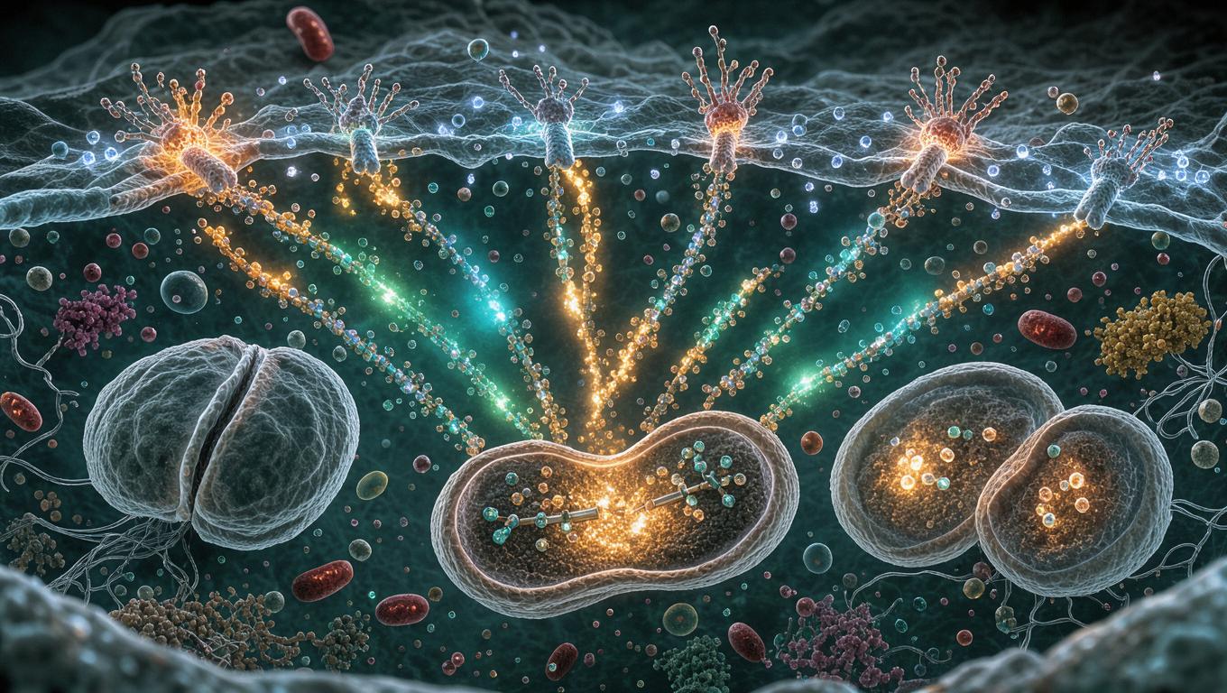
Sasaki et al. (eLife, 2025) show that mild membrane depolarization (15–30 mM extracellular K⁺, within near-physiological voltage range) activates ERK and enhances mitotic activity in human cell lines, with phosphatidylserine nanoclustering and Ras activation as upstream events The mechanistic chain is well-supported by imaging kinetics, but causality rests heavily on fendiline-based PS depletion, and generalization is limited to transformed cell lines in vitro.
Long Explanation
Core Evidence Chain
The paper links three layers: voltage → phosphatidylserine/Ras → ERK → mitosis. Depolarization of U2OS cells with 15 mM K⁺ increased cytokinesis (p=0.016), an effect abolished by the MEK inhibitor U0126 (p=0.0040), placing MEK-ERK causally upstream . FRET imaging (EKAREV) revealed graded ERK activation: near-maximal at 145 mM K⁺, ~50–60% at 30 mM, and heterogeneous with non-responders at 15 mM (Pearson r=0.54 vs log[K⁺]). Critically, perforated patch-clamp confirmed voltage itself, not secondary effects, drives ERK: −40 mV failed, −20 mV activated, and 0 mV reached ~88 ± 35% of EGF-maximal activity, with ERK deactivating within ~1 min of repolarization . FLIM-FRET showed phosphatidylserine nanoclustering and Ras activation within ~2 min of depolarization, preceding ERK, and fendiline-mediated PS depletion markedly blunted ERK activation (p=4.4×10⁻⁹) .
Note: values for 30K, 15K, and −20 mV are approximate ranges stated in the paper text, not exact figure-derived numbers.
Strengths and Cautions
Strengths: Combining single-cell FRET kinetics with voltage-clamp electrophysiology is a genuinely rigorous design; the patch-clamp data exclude perfusion artifacts and show ERK acts as an analog, reversible voltage readout rather than an on/off switch. Multi-line replication (U2OS, HEK293T, HeLa, A431) and proper controls (perfusion-only, hypotonic shock, Ca²⁺-free) strengthen the claim .
Cautions (BGPT critical appraisal): (1) The authors themselves note reliance on the Nernst equation for perfusion-based estimates — a partially addressed limitation, mitigated only by the HEK293T patch data, which cover a narrow voltage range (−80 to 0 mV in one cell type). (2) Fendiline is not PS-specific (it inhibits acid sphingomyelinase and can have off-target Ca²⁺ channel effects), so the PS-necessity claim would benefit from genetic PS-synthesis knockdown. (3) Single-cell n per condition is small (15–23 cells, 2–6 experiments), and the claim of a "universal" mechanism rests on four cancer-derived lines — non-transformed primary cells are untested. (4) Mitosis data show depolarization and FBS effects are non-additive, suggesting a plateau; whether physiological depolarization events in vivo reach the required thresholds is unknown. (5) The claimed link to Zhou et al.'s PS/K-Ras framework is correlative here; direct visualization of K-Ras relocalization in this system is absent.
What would falsify the model: PS-synthesis-conditional cells showing intact voltage-gated ERK activation despite PS depletion; non-transformed primary cells lacking voltage-dependent ERK activation; or failure of ERK activity to track voltage in a broader panel of resting potentials.
Feedback:
Updated: October 27, 2026
BGPT Paper Review
Study Novelty
70%
Extends Zhou et al. 2015 (PS/K-Ras voltage dependence) into the physiological voltage range and links it causally to mitosis — a meaningful but incremental advance over an existing mechanistic framework.
Scientific Quality
80%
Methodologically strong (FRET + patch clamp, proper controls, STR-authenticated cells), but fendiline specificity is imperfect, single-cell n is modest (15–23), and only cancer lines were tested. No prompt injections detected.
Study Generality
70%
The voltage-PS-Ras-ERK axis is plausibly universal to mammalian cells and connects bioelectricity to a core signaling pathway, but evidence is confined to four in vitro cancer-derived lines.
Study Usefulness
80%
Provides a mechanistic bridge between bioelectricity and proliferation with direct implications for cancer targeting (ion channels/transporters as upstream ERK regulators) and reinterprets stochastic ERK pulses.
Study Reproducibility
80%
Detailed methods, source blots, defined solutions and instruments; however, single-cell ERK variability and perfusion/pipette-dependent setups require careful replication.
We'll email you the results when your analysis is finished.
Hypothesis Graveyard
'Voltage-gated Ca²⁺ influx mediates depolarization-induced ERK activation' — directly falsified by the paper's Ca²⁺-free experiments, which preserved full ERK activation.
'ERK activation requires >50 mM extracellular K⁺ with wild-type K-Ras (Zhou et al. 2015)' — contradicted by single-cell FRET detection of ERK activation at 15–30 mM K⁺; the discrepancy likely reflects western blot averaging masking single-cell responses.

 Explore by Goal
 Quick Explanation
 Long Explanation
 Top Data Sources
 Ask a Follow-Up
 Key Insight
 Keep Exploring
 Hypothesis Graveyard
 Potential Experiments
 Discussion
