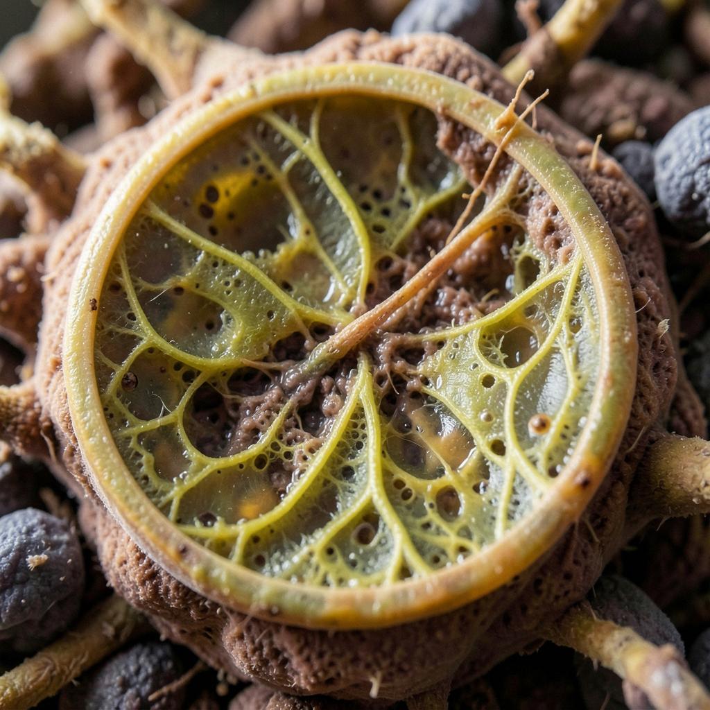
The Sucre lab (Vanderbilt) reports that postnatal alveoli form by epithelial clustering and directional extrusion through ring-like Pdgfra+ mesenchymal structures, followed by AT2βAT1 flatteningβnot by ingrowing septa. Key evidence: (1) 2,200 hours of SOPi volumetric live imaging found no dynamically subdividing septa; (2) apparent 2D "septal tips" resolve as parts of 3D rings when z-stacks are rendered; (3) epithelial somata displace 60β100 Β΅m through rings, matching normal P5 alveolar diameter, suggesting self-organizing sizing; (4) ML-7 (MLCK inhibition) arrests ring contraction and epithelial movement; (5) both Wnt activation (CHIR) and inhibition (XAV-939) abolish alveologenesis events and PDPN+ differentiation .
Graph note: qualitative illustration of ex vivo fidelity claims; the paper reports histologic/SEM equivalence at 48h but explicitly states long ex vivo culture does not reproduce in vivo ring loss by P14.
In vivo intravital imaging or serial 3D reconstruction of unsectioned lungs showing septal ingrowth as a major contributor; or demonstration that alveologenesis proceeds normally when ring contraction is genetically (not pharmacologically) abolished with epithelial extrusion intact.
Know what changed, what holds up, and what remains uncertain. Every Friday. No ads.