Why BGPT?
logo

Paper Review — Verify Claims Fast

Quickly check methods, data, and figures across full-text papers to verify conclusions.

Press Enter ↵ to review


    Fuel Your Discoveries


     Quick Explanation


    Core claim check (from the provided paper text): the study proposes a causal cascade where Aβ exposure activates astrocytic NADPH oxidase → oxidative stress → PARP activation and PAR accumulation → NAD+/NADH redox disruption and mitochondrial substrate limitation → mitochondrial membrane potential loss → (primarily) neuronal death, with PARP inhibition and NADPH oxidase inhibition preventing key steps and toxicity.


     Long Explanation


    Paper Review (Science-Critical): β-amyloid → PARP → astrocytic metabolic failure → neuronal death

    1) Visual mechanism map (what the authors argue)

    Evidence grounding: This pathway diagram reflects the paper’s abstract + Results/Discussion narrative: Aβ activates astrocytic NADPH oxidase, increases PAR in astrocytes, decreases NADH and oxygen consumption, and PARP inhibitors prevent PAR and mitochondrial depolarization; co-culture neuronal death after 48 h is prevented by PARP inhibition.

    2) Data extraction visualizations (from the provided raw data list)

    Methodological humility on the visuals: The above plots use only the numeric values present in the user-provided “RESEARCH DATA TO UTILIZE + GRAPH” list; the full paper contains additional time points, replicates, and inhibitor-specific comparisons that are not numerically included in that list.

    3) Step-by-step critique of the causal chain

    A) Aβ → astrocytic NADPH oxidase activation → oxidative stress

    • What’s claimed: Aβ exposure increases ROS generation via the glial NADPH oxidase, and inhibiting/knocking down NADPH oxidase prevents mitochondrial depolarization phenotypes.
    • Key skeptical check: The paper relies on pharmacological inhibitors (e.g., diphenylene iodonium) and interpretation that this specifically maps to NADPH oxidase. Without independent, direct ROS flux quantification at matched timing/locations for each inhibitor condition in the excerpt you provided, some uncertainty remains about off-target contributions.

    B) Oxidative stress → PARP activation → PAR accumulation in astrocytes

    • What’s claimed: Aβ increases PAR polymers in GFAP+ astrocytes within ~1 h, and this PAR accumulation is prevented by PARP inhibition and by NADPH oxidase inhibition.
    • What is still uncertain: PAR polymer immunofluorescence is an informative readout, but the excerpt does not show whether PAR localization/turnover kinetics, assay linearity, and PAR species specificity were benchmarked (e.g., via orthogonal enzymatic PARP activity assays).

    C) PARP activation → NAD+ depletion / NADH signal changes + oxygen consumption drop

    • What’s claimed: Aβ decreases NADH autofluorescence and reduces oxygen consumption; mitochondrial substrates reverse the mitochondrial depolarization, consistent with substrate limitation rather than primarily direct electron-transport chain damage.
    • Mechanistic inference to flag: NADH autofluorescence can be influenced by multiple factors (probe localization, redox state, imaging calibration). The paper notes fura-2 calcium data were not calibrated for [Ca2+]c due to calibration uncertainty; it does not, in the provided excerpt, quantify uncertainty in NADH signal interpretation.

    D) Mitochondrial depolarization phenotype: PARP-dependent “slow” vs cyclophilin D–dependent “transient” events

    • What’s claimed: b-amyloid causes two components in astrocytes: (i) ROS-dependent slow progressive depolarization, and (ii) Ca2+- and ROS-dependent transient depolarizations prevented by cyclophilin D knockout (mitochondrial permeability transition pore regulator). PARP inhibitors block the slow depolarization and also block the slow response induced by PMA.
    • Critical nuance: In the Discussion, the authors argue that reversible permeability transition pore events play a “very small” role in cell death in their model, based on observations that reducing/altering permeability transition can accelerate the slow depolarization. That is a non-trivial inference and could be sensitive to how “cell death” endpoints track with mitochondrial potential dynamics.

    E) PARP inhibition protects neurons in co-culture and in aged transgenic mice

    • What’s claimed: In neuron/astrocyte co-cultures exposed to Aβ for ~24–48 h, PARP inhibition reduces neuronal death and prevents NADH depletion and mitochondrial membrane potential loss; PAR polymers accumulate in vivo in the TASTPM Alzheimer model with increasing age.
    • Translational blind spot: The in vitro concentrations and peptide forms (Aβ1-42 and fragment 25-35) are not guaranteed to match physiological aggregation states, receptor engagements, or kinetics in human disease. The paper does attempt to use molar excess arguments for peptide presence vs inhibitors and notes that reverse peptide controls were negative, but physiological relevance remains an uncertainty.

    4) Relation to other Aβ oxidative-stress mechanisms (context, not proof)

    Cross-check example: Another study in the provided dataset links Aβ25-35–induced neuronal death to neuronal NADPH oxidase (NOX) activation and gp91phox knockdown reducing ROS and death in mixed cortical cultures. This partially overlaps with the current paper’s NOX→ROS→death concept but does not directly validate the PARP–NAD+–mitochondrial substrate axis.
    What this means for your paper: it supports the broader plausibility that NOX activation can mediate Aβ neurotoxicity in culture, but it does not confirm that PARP is the critical downstream node in all models.

    5) What would most convincingly falsify the paper’s main mechanistic claim?

    • PARP independence: If Aβ exposure produces the mitochondrial depolarization and neuronal death phenotypes even when PARP activity is equivalently suppressed (with multiple orthogonal PARP inhibition strategies that independently block PAR-dependent PAR polymer accumulation).
    • NOX independence: If Aβ toxicity proceeds without detectable astrocytic NOX-driven oxidative stress (e.g., NOX inhibition fails to prevent PAR polymer accumulation and mitochondrial redox/substrate phenotypes).
    • Alternative substrate-failure mechanism: If mitochondrial depolarization and oxygen consumption changes are reproduced with PARP inhibition alone (i.e., not restored by restoring substrate supply or NAD+/redox correction), undermining the claim that PARP consumes NAD+ and thereby limits substrate availability.

    6) Quick decision table: claims vs evidence type vs confidence

    Claim Evidence in provided text Evidence strength Key uncertainty / blind spot
    Aβ activates astrocytic NADPH oxidase and drives oxidative stress NOX inhibition prevents mitochondrial depolarization + neuronal toxicity; abstract describes NOX activation strong inhibitor specificity/OFF-target considerations not fully resolved in excerpt
    Aβ → PAR accumulation specifically in astrocytes anti-PAR immunofluorescence; decreased by NADPH oxidase inhibition and PARP inhibition strong orthogonal PARP activity assays not shown in excerpt
    PARP activation depletes NAD+ and limits mitochondrial substrate supply NADH autofluorescence↓ + oxygen consumption↓; substrate provision reverses depolarization; PARP inhibitors prevent slow depolarization moderate–strong NADH imaging-to-pool quantification assumptions
    Neuronal death in co-culture depends on PARP in response to astrocytic NOX/ROS neuronal death and neuronal mitochondrial/NADH changes prevented by PARP inhibitors strong in vitro model relevance to human disease microenvironment
    PAR polymer accumulation increases with age in TASTPM mice in vivo immunofluorescence for PAR polymers in brain sections moderate correlation with disease stage; does not prove mechanistic causality in vivo from this excerpt alone

    Run Science AI Agent (optional)


    Feedback:   

    Updated: April 13, 2026

    BGPT Paper Review


    Study Novelty

    70%

    The mechanistic framing (astrocytic NOX→oxidative stress→PARP activation→NAD+ depletion→mitochondrial substrate limitation→neuron death) is a specific, testable causal sequence rather than a generic association; however, it builds on well-known roles of PARP/NAD+ in oxidative-stress and cell death biology, reducing “completely new” novelty.


    Scientific Quality

    80%

    Strengths: multi-level readouts (mitochondrial potential, NAD(P)H autofluorescence, oxygen consumption, PAR immunofluorescence, cell death), genetic dissection (cyclophilin D knockout to separate transient depolarizations), and pharmacological prevention (NOX/PARP inhibitors). Main limitations to flag from the provided excerpt: reliance on inhibitor-based specificity and an in vitro peptide-exposure paradigm with uncertain physiological aggregation/state matching.


    Study Generality

    60%

    The model is fairly specific: mixed hippocampal neuron–glia co-cultures, particular Aβ fragments/concentrations, and a PARP–mitochondrial substrate limitation mechanism in astrocytes. While the upstream logic (oxidative stress→PARP→NAD+ depletion→mitochondrial dysfunction) may generalize to other oxidative insults, the exact mapping to human AD pathophysiology remains uncertain.


    Study Usefulness

    70%

    Usefulness is high for hypothesis generation and mechanistic targeting: it offers a structured chain with measurable intermediates (PAR accumulation, NADH/redox, oxygen consumption, mitochondrial potential) and clear inhibitor-rescue logic, but translational applicability depends on whether the same node dominance holds in human brain and across Aβ species/states.


    Study Reproducibility

    60%

    Many experimental details are present (cell culture source/age, inhibitors, imaging/oxygen electrode methods), but reproducibility is reduced by reliance on specialized primary co-culture conditions, peptide handling at high concentrations, and PARP/NOX inhibitor choice where off-targets and assay variability can matter.


    Explanatory Depth

    80%

    The paper provides a mechanistic sequence with mechanistic separation (cyclophilin D knockout disentangles transient permeability transition events from the PARP-dependent slow component) and proposes how PARP-linked NAD+ depletion could lead to impaired substrate supply and mitochondrial redox/respiration changes.


    🎁 Authors: Collect 225 Free Science Tokens (≈ $22.5 USD)

    Claim My Author Tokens

    Use for 56 days of free BGPT access (4 tokens = 1 day) or trade/sell (≈ $22.5 USD)

     Top Data Sources ExportMCP


     Analysis Wizard


    Extracts the provided numeric summaries (NADH decrease, O2 consumption baseline vs Aβ, neuron vs astrocyte death) into tidy arrays and generates Plotly-ready bar charts for rapid comparison across endpoints.


     Hypothesis Graveyard


    “Mitochondrial permeability transition pore transients are the primary trigger for neuronal death in this model.” The paper argues these transients are largely separable and that death depends mainly on slow PARP-linked substrate/NAD+ depletion; thus this hypothesis is already undermined by their cyclophilin D separation logic (though with some interpretive uncertainty).


    “PAR polymer accumulation is epiphenomenal and does not map onto NAD+/mitochondrial substrate limitation.” This is weakened by the paper’s inhibitor-rescue results showing PARP inhibition prevents both PAR accumulation and NADH/respiration/mitochondrial depolarization phenotypes.

     Science Art


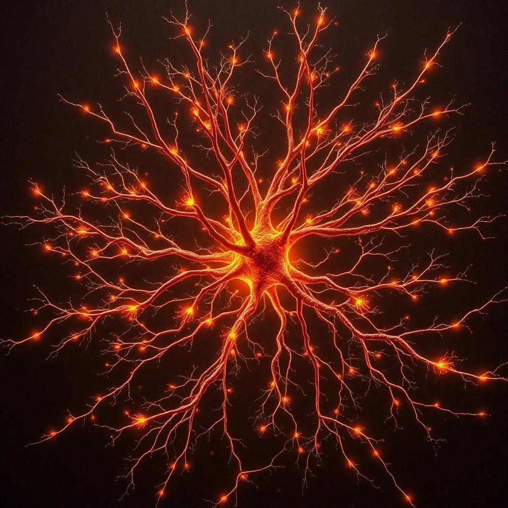
    Paper Review: β-amyloid activates PARP causing astrocytic metabolic failure and neuronal death Science Art

     Science Movie


    Make a narrated HD Science movie for this answer ($32 per minute)


     Discussion


    Get Ahead With Science Insights

    Custom summaries of the latest cutting-edge research. Every Friday. No ads.


    My BGPT